
 KSCRS

Articles
- Page Path
- HOME > Insights Cataract Refract Surg > Volume 11(3); 2026 > Article
-
Original Article
건조증이 있는 녹내장 환자에서 시야검사 전 인공눈물의 효과 -
이지영, 김은철

-
Effect of lubricant eye drops administered before visual field testing in patients with glaucoma and dry eye -
Jiyoung Emily Lee, Eun Chul Kim
-
Insights in Cataract and Refractive Surgery 2026;11(3):91-97.
DOI: https://doi.org/10.63375/icrs.26.009
Published online: October 29, 2026
가톨릭대학교 의과대학 안과학교실, 부천성모병원
Department of Ophthalmology, Bucheon St. Mary’s Hospital, College of Medicine, The Catholic University of Korea, Seoul, Korea
- Correspondence to: Eun Chul Kim Department of Ophthalmology, Bucheon St. Mary’s Hospital, 327 Sosa-ro, Wonmi-gu, Bucheon 14647, Korea Tel: +82-32-340-2125 E-mail: eunchol@hanmail.net
• Received: August 20, 2026 • Revised: September 24, 2026 • Accepted: September 29, 2026
© 2026 Korean Society of Cataract and Refractive Surgery.
This is an Open Access article distributed under the terms of the Creative Commons Attribution Non-Commercial License (https://creativecommons.org/licenses/by-nc/4.0/) which permits unrestricted non-commercial use, distribution, and reproduction in any medium, provided the original work is properly cited.
- 37 Views
- 0 Download


국문초록
-
목적
- 안구건조증이 있는 녹내장 환자에서 시야검사 전 인공눈물 점안에 따른 효과를 분석하고자 하였다.
-
방법
- 안구건조증 환자 30명, 60안을 대상으로 시야검사 시행 전 우안은 히알루론산 0.1% 안약을 점안하였고, 좌안은 점안 없이 시행하였다. 시야검사 시행 전 후 눈물높이(tear height), 비침습눈물막파괴시간(NiBUT), 각막염색검사(staining score)의 변화를 후향적으로 분석하여 인공눈물의 효과를 분석하였다.
-
결과
- 시야검사 전 비침습눈물막파괴시간은 우안 5.16±2.19초, 좌안 5.07±1.98초, 시야검사 후의 경우 우안 5.04±2.36초, 좌안 4.27±1.32초 측정되었고, 눈물높이의 경우 시야검사 전 우안 0.26±0.11 mm, 좌안 0.25±0.10 mm, 시야검사 후 우안 0.25±0.11 mm 좌안 0.22±0.10 mm가 측정되었으며, 각막염색검사의 경우 시야검사 전 우안 1.07±0.74, 좌안 1.17±0.86, 우안 1.33±0.76, 좌안 1.90±0.95이었다. 시야검사 전과 비교하여 시야검사 후 우안보다 좌안의 비침습눈물파괴시간과 눈물높이가 각각 감소되었고 각막염색이 유의하게 증가하였다(P<0.05). 시야검사의 결과는 좌안에서 주시실패율, 위양성률, 위음성률이 우안보다 각각 유의하게 높았다(P<0.05).
-
결론
- 안구건조증 환자들의 경우 오랜 시간 동안 눈을 뜨고 집중해야 하는 시야검사로 건조증의 징후가 악화되며 시야검사의 정확도도 낮아질 수 있다. 안과전문의의 진료를 받은 후 인공눈물을 점안하고 시야검사를 시행하면 안구건조증의 징후를 완화시키며 더 정확한 시야검사가 될 것으로 사료된다.
Abstract
-
Purpose
- This study aimed to evaluate the effect of administering lubricant eye drops before visual field testing in patients with glaucoma and dry eye syndrome.
-
Methods
- The records of 30 patients (60 eyes) with dry eye syndrome were retrospectively reviewed. Before visual field testing, 0.1% hyaluronic acid eye drops were administered to the right eye, while the left eye received no drops. The efficacy of the lubricant eye drops was assessed by retrospectively comparing tear meniscus height, noninvasive tear film break-up time (NiBUT), and corneal staining scores before and after testing.
-
Results
- Before visual field testing, NiBUT was 5.16±2.19 seconds in the right eye and 5.07±1.98 seconds in the left eye; after testing, it was 5.04±2.36 seconds and 4.27±1.32 seconds, respectively. Tear meniscus height was 0.26±0.11 mm in the right eye and 0.25±0.10 mm in the left eye before testing, decreasing to 0.25±0.11 mm and 0.22±0.10 mm, respectively, afterward. Before testing, corneal staining scores were 1.07±0.74 in the right eye and 1.17±0.86 in the left eye; after testing, they were 1.33±0.76 and 1.90±0.95, respectively. The decreases in NiBUT and tear meniscus height from before to after testing were greater in the left eye than in the right eye, and corneal staining increased significantly (P<0.05). Fixation loss, false-positive, and false-negative rates during visual field testing were all significantly higher in the left eye than in the right eye (P<0.05).
-
Conclusion
- Visual field testing requires patients to keep their eyes open and maintain focus for an extended period, which can exacerbate symptoms in patients with dry eye syndrome and potentially compromise test accuracy. Administering lubricant eye drops before testing, following consultation with an ophthalmologist, is thought to alleviate dry eye symptoms and improve test accuracy.
서론
방법
결과
고찰
Article Information
Author contributions
Conceptualization, JEL, ECK. Data curation,JEL, ECK. Funding acquisition, ECK. Investigation, JEL, ECK. Methodology, JEL, ECK. Project administration, JEL, ECK. Supervision, ECK. Formal analysis, JEL, ECK. Writing_original draft, JEL, ECK. Writing_review & editing, ECK.
Conflicts of interest
No potential conflict of interest relevant to this article was reported.
Funding
None.
Data availability
The data that support the findings of this study are available from the corresponding author upon reasonable request.
Fig. 1.Change in tear meniscus height after visual field testing. HA, hyaluronic acid; Rt, right; Lt, left. The decrease in tear meniscus height from before to after visual field testing was greater in the left eye than in the right eye (P<0.05). The right and left eyes were compared using the Mann–Whitney test. Error bars represent standard deviation. *P<0.05.

Fig. 2.Change in noninvasive tear film break-up time (NiBUT) after visual field testing. HA, hyaluronic acid; Rt, right; Lt, left. The decrease in NiBUT from before to after visual field testing was greater in the left eye than in the right eye (P<0.05). The right and left eyes were compared using the Mann–Whitney test. Error bars represent standard deviation. *P<0.05.

Fig. 3.Change in corneal fluorescein staining grade after visual field testing. HA, hyaluronic acid; Rt, right; Lt, left. The increase in corneal fluorescein staining grade from before to after visual field testing was greater in the left eye than in the right eye (P<0.05). The right and left eyes were compared using the Mann–Whitney test. Error bars represent standard deviation. *P<0.05.

Table 1.Patient characteristics (n=30)
Table 2.Dry eye signs before and after visual field testing
| Variable | Right eye (0.1% HA) (n=30) | Left eye (n=30) |
|---|---|---|
| Schirmer I test | 5.45±2.69 | 5.78±3.01 |
| Tear meniscus height (mm) | ||
| Before visual field testing | 0.26±0.11 | 0.25±0.10 |
| After visual field testing | 0.25±0.11 | 0.22±0.10* |
| NiBUT (sec) | ||
| Before visual field testing | 5.16±2.19 | 5.07±1.98 |
| After visual field testing | 5.04±2.36 | 4.27±1.32* |
| Corneal staining grade | ||
| Before visual field testing | 1.07±0.74 | 1.17±0.86 |
| After visual field testing | 1.33±0.76 | 1.90±0.95* |
Values are presented as mean±standard deviation.
NiBUT, noninvasive tear film break-up time; HA, hyaluronic acid.
Before visual field testing, none of the parameters differed significantly between the right and left eyes. After testing, noninvasive tear film break-up time and tear meniscus height were lower and corneal staining was higher in the left eye than in the right eye (P<0.05). The right and left eyes were compared using the Mann–Whitney test.
*P<0.05.
Table 3.Visual field test results
| Variable | Right eye (0.1% HA) (n=30) | Left eye (n=30) |
|---|---|---|
| Fixation losses (%) | 20.66±7.41 | 26.63±8.61* |
| False-positive errors (%) | 2.47±1.26 | 7.01±2.55* |
| False-negative errors (%) | 2.80±1.08 | 5.67±2.31* |
| Mean deviation | -4.68±3.82 | -4.25±3.59 |
| Pattern standard deviation | 3.85±1.79 | 3.79±1.58 |
- 1. Pouyeh B, Viteri E, Feuer W, et al. Impact of ocular surface symptoms on quality of life in a United States veterans affairs population. Am J Ophthalmol 2012;153:1061‒6.e3. ArticlePubMed
- 2. Karakus S, Mathews PM, Agrawal D, et al. Impact of dry eye on prolonged reading. Optom Vis Sci 2018;95:1105‒13. ArticlePubMedPDF
- 3. Herbaut A, Liang H, Denoyer A, et al. Tear film analysis and evaluation of optical quality: a review of the literature. J Fr Ophtalmol 2019;42:e21‒e35. ArticlePubMed
- 4. Koh S. Mechanisms of visual disturbance in dry eye. Cornea 2016;35 Suppl 1:S83‒S88. ArticlePubMed
- 5. Tirpack AR, Vanner E, Parrish JM, et al. Dry eye symptoms and ocular pain in veterans with glaucoma. J Clin Med 2019;8:1076.ArticlePubMedPMC
- 6. Walsh K, Jones L. The use of preservatives in dry eye drops. Clin Ophthalmol 2019;13:1409‒25. ArticlePubMedPMC
- 7. Ramli N, Supramaniam G, Samsudin A, et al. Ocular surface disease in glaucoma: effect of polypharmacy and preservatives. Optom Vis Sci 2015;92:e222‒e226. ArticlePubMed
- 8. Brandt JD, Wittpenn JR, Katz LJ, et al. Conjunctival impression cytology in patients with glaucoma using long-term topical medication. Am J Ophthalmol 1991;112:297‒301. ArticlePubMed
- 9. Huang FC, Tseng SH, Shih MH, Chen FK. Effect of artificial tears on corneal surface regularity, contrast sensitivity, and glare disability in dry eyes. Ophthalmology 2002;109:1934‒40. ArticlePubMed
- 10. Rieger G. Contrast sensitivity in patients with keratoconjunctivitis sicca before and after artificial tear application. Graefes Arch Clin Exp Ophthalmol 1993;231:577‒9. ArticlePubMedPDF
- 11. Novak KD, Kohnen T, Chang-Godinich A, et al. Changes in computerized videokeratography induced by artificial tears. J Cataract Refract Surg 1997;23:1023‒8. ArticlePubMed
- 12. Miranda MA, Henson DB. Perimetric sensitivity and response variability in glaucoma with single-stimulus automated perimetry and multiple-stimulus perimetry with verbal feedback. Acta Ophthalmol 2008;86:202‒6. ArticlePubMed
- 13. McMillan TA, Stewart WC, Hunt HH. Association of reliability with reproducibility of the glaucomatous visual field. Acta Ophthalmol (Copenh) 1992;70:665‒70. ArticlePubMed
- 14. Ishiyama Y, Murata H, Mayama C, Asaoka R. An objective evaluation of gaze tracking in Humphrey perimetry and the relation with the reproducibility of visual fields: a pilot study in glaucoma. Invest Ophthalmol Vis Sci 2014;55:8149‒52. ArticlePubMed
- 15. Ishiyama Y, Murata H, Asaoka R. The usefulness of gaze tracking as an index of visual field reliability in glaucoma patients. Invest Ophthalmol Vis Sci 2015;56:6233‒6. ArticlePubMed
- 16. Rieger G. The importance of the precorneal tear film for the quality of optical imaging. Br J Ophthalmol 1992;76:157‒8. ArticlePubMedPMC
- 17. Herse PR. Factors influencing normal perimetric thresholds obtained using the Humphrey Field Analyzer. Invest Ophthalmol Vis Sci 1992;33:611‒7. PubMed
- 18. Sagara H, Sekiryu T, Imaizumi K, et al. Impact of tear metrics on the reliability of perimetry in patients with dry eye. PLoS One 2019;14:e0222467. ArticlePubMedPMC
- 19. Arai T, Murata H, Matsuura M, et al. The association between ocular surface measurements with visual field reliability indices and gaze tracking results in preperimetric glaucoma. Br J Ophthalmol 2018;102:525‒30. ArticlePubMed
- 20. Herreras JM, Pastor JC, Calonge M, Asensio VM. Ocular surface alteration after long-term treatment with an antiglaucomatous drug. Ophthalmology 1992;99:1082‒8. ArticlePubMed
- 21. De Saint Jean M, Debbasch C, Brignole F, et al. Toxicity of preserved and unpreserved antiglaucoma topical drugs in an in vitro model of conjunctival cells. Curr Eye Res 2000;20:85‒94. ArticlePubMed
- 22. Yenice O, Temel A, Orum O. The effect of artificial tear administration on visual field testing in patients with glaucoma and dry eye. Eye (Lond) 2007;21:214‒7. ArticlePubMedPDF
- 23. Ozyol P, Ozyol E, Karalezli A. Evaluation of visual field test parameters after artificial tear administration in patients with glaucoma and dry eye. Semin Ophthalmol 2018;33:320‒4. ArticlePubMed
- 24. Guzey M, Satici A, Karaman SK, et al. The effect of lubricating eye drop containing hydroxypropyl guar on perimetry results of patients with glaucoma and trachomatous dry eye. Ophthalmologica 2010;224:109‒15. ArticlePubMedPDF
- 25. Kocabeyoglu S, Mocan MC, Bozkurt B, Irkec M. Effect of artificial tears on automated visual field testing in patients with glaucoma and dry eye. Can J Ophthalmol 2013;48:110‒4. ArticlePubMed
- 26. Heijl A, Patella VM. Essential perimetry: the field analyzer primer. Jena, Germany: Carl Zeiss Meditec Inc.; 2002.
References
Figure & Data
References
Citations
Citations to this article as recorded by 
 ePub Link
 Cite
 XML Download
Effect of lubricant eye drops administered before visual field testing in patients with glaucoma and dry eye
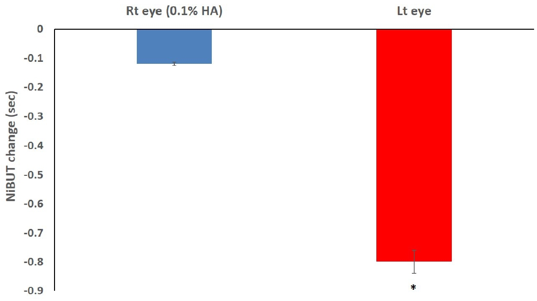


Fig. 1. Change in tear meniscus height after visual field testing. HA, hyaluronic acid; Rt, right; Lt, left. The decrease in tear meniscus height from before to after visual field testing was greater in the left eye than in the right eye (P<0.05). The right and left eyes were compared using the Mann–Whitney test. Error bars represent standard deviation. *P<0.05.
Fig. 2. Change in noninvasive tear film break-up time (NiBUT) after visual field testing. HA, hyaluronic acid; Rt, right; Lt, left. The decrease in NiBUT from before to after visual field testing was greater in the left eye than in the right eye (P<0.05). The right and left eyes were compared using the Mann–Whitney test. Error bars represent standard deviation. *P<0.05.
Fig. 3. Change in corneal fluorescein staining grade after visual field testing. HA, hyaluronic acid; Rt, right; Lt, left. The increase in corneal fluorescein staining grade from before to after visual field testing was greater in the left eye than in the right eye (P<0.05). The right and left eyes were compared using the Mann–Whitney test. Error bars represent standard deviation. *P<0.05.
Fig. 1.
Fig. 2.
Fig. 3.
Effect of lubricant eye drops administered before visual field testing in patients with glaucoma and dry eye
| Characteristic | Value |
|---|---|
| Age (yr) | 67.9±9.7 |
| Sex | |
| Male | 12 |
| Female | 18 |
| Duration of glaucoma (yr) | 7.5±4.8 |
| Glaucoma medications | |
| Prostaglandin monotherapy | 12 (40) |
| Prostaglandin/beta-blocker combination | 8 (26.7) |
| Dorzolamide/beta-blocker combination | 10 (33.3) |
| Variable | Right eye (0.1% HA) (n=30) | Left eye (n=30) |
|---|---|---|
| Schirmer I test | 5.45±2.69 | 5.78±3.01 |
| Tear meniscus height (mm) | ||
| Before visual field testing | 0.26±0.11 | 0.25±0.10 |
| After visual field testing | 0.25±0.11 | 0.22±0.10 |
| NiBUT (sec) | ||
| Before visual field testing | 5.16±2.19 | 5.07±1.98 |
| After visual field testing | 5.04±2.36 | 4.27±1.32 |
| Corneal staining grade | ||
| Before visual field testing | 1.07±0.74 | 1.17±0.86 |
| After visual field testing | 1.33±0.76 | 1.90±0.95 |
| Variable | Right eye (0.1% HA) (n=30) | Left eye (n=30) |
|---|---|---|
| Fixation losses (%) | 20.66±7.41 | 26.63±8.61 |
| False-positive errors (%) | 2.47±1.26 | 7.01±2.55 |
| False-negative errors (%) | 2.80±1.08 | 5.67±2.31 |
| Mean deviation | -4.68±3.82 | -4.25±3.59 |
| Pattern standard deviation | 3.85±1.79 | 3.79±1.58 |
Table 1. Patient characteristics (n=30)
Values are presented as mean±standard deviation or number (%).
Table 2. Dry eye signs before and after visual field testing
Values are presented as mean±standard deviation. NiBUT, noninvasive tear film break-up time; HA, hyaluronic acid. Before visual field testing, none of the parameters differed significantly between the right and left eyes. After testing, noninvasive tear film break-up time and tear meniscus height were lower and corneal staining was higher in the left eye than in the right eye (P<0.05). The right and left eyes were compared using the Mann–Whitney test. P<0.05.
Table 3. Visual field test results
Values are presented as mean±standard deviation. HA, hyaluronic acid. Fixation losses, false-positive errors, and false-negative errors were significantly higher in the left eye than in the right eye (P<0.05). The right and left eyes were compared using the Mann–Whitney test. P<0.05.


