

Skip Navigation
Skip to contents

Insights Cataract Refract Surg : Insights in Cataract and Refractive Surgery

OPEN ACCESS

Articles

Page Path
HOME > Insights Cataract Refract Surg > Ahead-of print articles > Article
Case Report
Therapeutic penetrating keratoplasty in rapidly progressive Acanthamoeba keratitis: a case report
Yunseo Choi1,*, Jaewon Jung1,*, Tae-im Kim2,3orcid

DOI: https://doi.org/10.63375/icrs.26.006
Published online: July 31, 2026

1Yonsei University College of Medicine, Seoul, Korea

2The Institute of Vision Research, Department of Ophthalmology, Yonsei University College of Medicine, Seoul, Korea

3Corneal Dystrophy Research Institute, Department of Ophthalmology, Yonsei University College of Medicine, Seoul, Korea

Correspondence to: Tae-im Kim Corneal Dystrophy Research Institute, Department of Ophthalmology, Yonsei University College of Medicine, 50 Yonsei-ro, Seodaemun-gu, Seoul 03722, Korea Tel: +82-2-2228-3592 E-mail: tikim@yuhs.ac
*Yunseo Choi and Jaewon Jung contributed equally to this work as co-first authors.
• Received: May 24, 2026   • Revised: June 10, 2026   • Accepted: June 10, 2026

© 2026 Korean Society of Cataract and Refractive Surgery.

This is an Open Access article distributed under the terms of the Creative Commons Attribution Non-Commercial License (https://creativecommons.org/licenses/by-nc/4.0/) which permits unrestricted non-commercial use, distribution, and reproduction in any medium, provided the original work is properly cited.

  • 197 Views
  • 3 Download
  • Purpose
    To report the clinical course and postoperative management strategy for emergency therapeutic penetrating keratoplasty (PKP) in a patient with rapidly progressive Acanthamoeba keratitis.
  • Case summary
    A 23-year-old female patient with a history of long-term orthokeratology lens wear presented with Acanthamoeba keratitis in the right eye. The diagnosis had been established at a referring hospital based on characteristic clinical findings, and topical antiamoebic therapy was initiated, resulting in initial clinical improvement. After polyhexamethylene biguanide was discontinued, an epithelial defect recurred. Amniotic membrane transplantation with adjunctive topical corticosteroids was then performed, after which corneal infiltration and inflammation worsened. At presentation to our clinic, best-corrected visual acuity (BCVA) was light perception, and intraocular pressure was elevated to 34.1 mmHg. Slit-lamp biomicroscopic examination revealed extensive corneal infiltration accompanied by hypopyon. Emergency therapeutic PKP was performed the following day because of uncontrolled infection with severe stromal thinning. Postoperatively, intensive antiamoebic therapy was administered, and topical corticosteroids were initially withheld. Systemic immunosuppression was introduced 1 week postoperatively to manage early graft rejection. Management required careful balancing of immunosuppression for graft survival against the risk of recurrent Acanthamoeba infection. Extensive inferior corneal neovascularization was observed and required additional interventions, including subconjunctival bevacizumab injection. At 5 to 6 months postoperatively, the graft remained clear, with no evidence of recurrent infection, and BCVA had improved to 0.7.
  • Conclusion
    Timely emergency therapeutic PKP with careful postoperative management may serve as a vision-preserving strategy in patients with rapidly progressive Acanthamoeba keratitis.
Acanthamoeba keratitis is a rare but potentially vision-threatening corneal infection, most commonly associated with contact lens use, including orthokeratology lenses [1,2]. It is characterized by severe ocular pain and corneal nerve inflammation [3]. Clinical features frequently mimic those of herpetic or bacterial keratitis, often leading to delayed diagnosis and initiation of inappropriate treatment [4]. A characteristic stromal ring infiltrate, when present, serves as an important diagnostic feature, typically observed in more advanced stages of the disease [5].
Despite advances in medical therapy, including biguanides and diamidines, treatment remains challenging due to cyst formation and the organism’s persistence, frequently resulting in prolonged disease course and poor visual outcomes [5,6]. In cases of advanced disease with severe stromal involvement, surgical intervention such as therapeutic penetrating keratoplasty (PKP) may be required to control infection and preserve ocular integrity [6]. Here, we report a case of rapidly progressive Acanthamoeba keratitis with severe stromal involvement requiring emergency therapeutic PKP. The requirement for informed consent was waived.
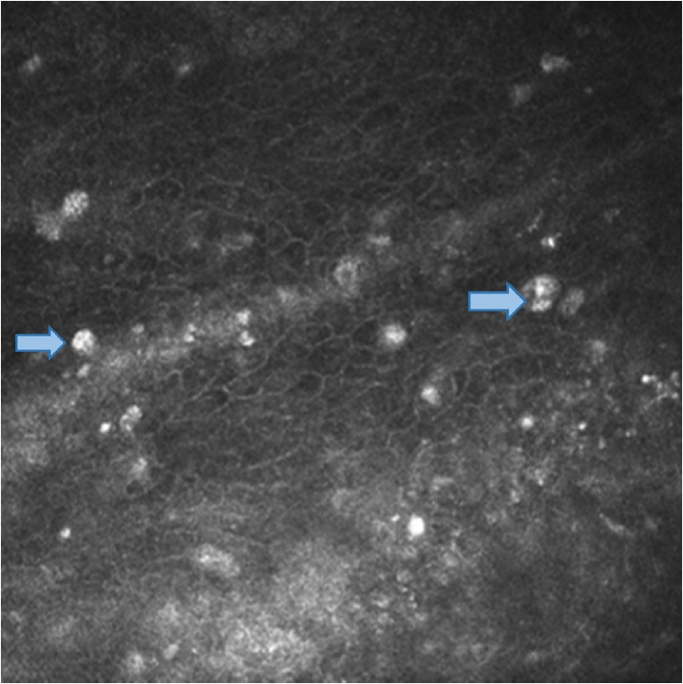
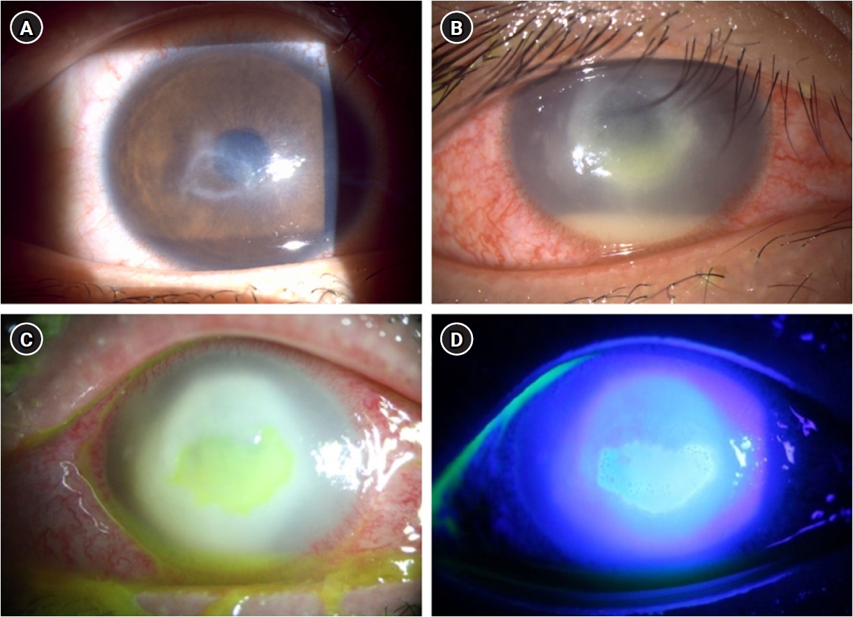
A 23-year-old female patient with no significant past medical history and a history of long-term orthokeratology lens wear presented with progressive visual loss and pain in the right eye. She had initially been evaluated at a university-based ophthalmology center in the United States approximately 3 months prior, where a clinical diagnosis of Acanthamoeba keratitis was established based on the characteristic stromal ring infiltrate, supported by hyperreflective cyst-like structures identified on in vivo confocal microscopy (Fig. 1). Topical antiamoebic therapy, including propamidine isethionate, polyhexamethylene biguanide (PHMB), and voriconazole, was initiated with improvement in corneal findings; PHMB was tapered and discontinued after 2 months of treatment. Corneal lesions recurred 2 weeks after discontinuation (Fig. 2A), and amniotic membrane transplantation was performed with concurrent antimicrobial and topical corticosteroid therapy. Despite these interventions, corneal infiltration and inflammation worsened within 1 week, and the patient was referred to our clinic for further management (Fig. 2B).
At initial presentation, best-corrected visual acuity (BCVA) was light perception in the right eye and 0.8 in the left eye. Biomicroscopy revealed extensive corneal infiltration with severe stromal thinning and a large epithelial defect, accompanied by hypopyon (Fig. 2C). Fluorescein staining revealed an epithelial defect involving the majority of the corneal surface (Fig. 2D). Intraocular pressure (IOP) was markedly elevated at 34.1 mmHg, measured by rebound tonometry (iCare, iCare Finland Oy), consistent with inflammatory trabecular compromise in the setting of severe anterior segment inflammation.
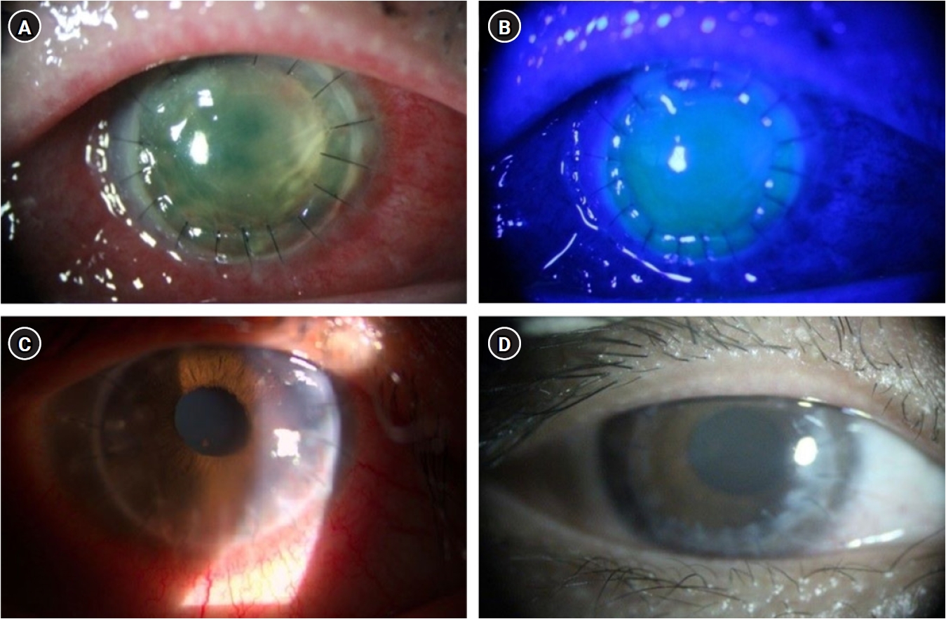
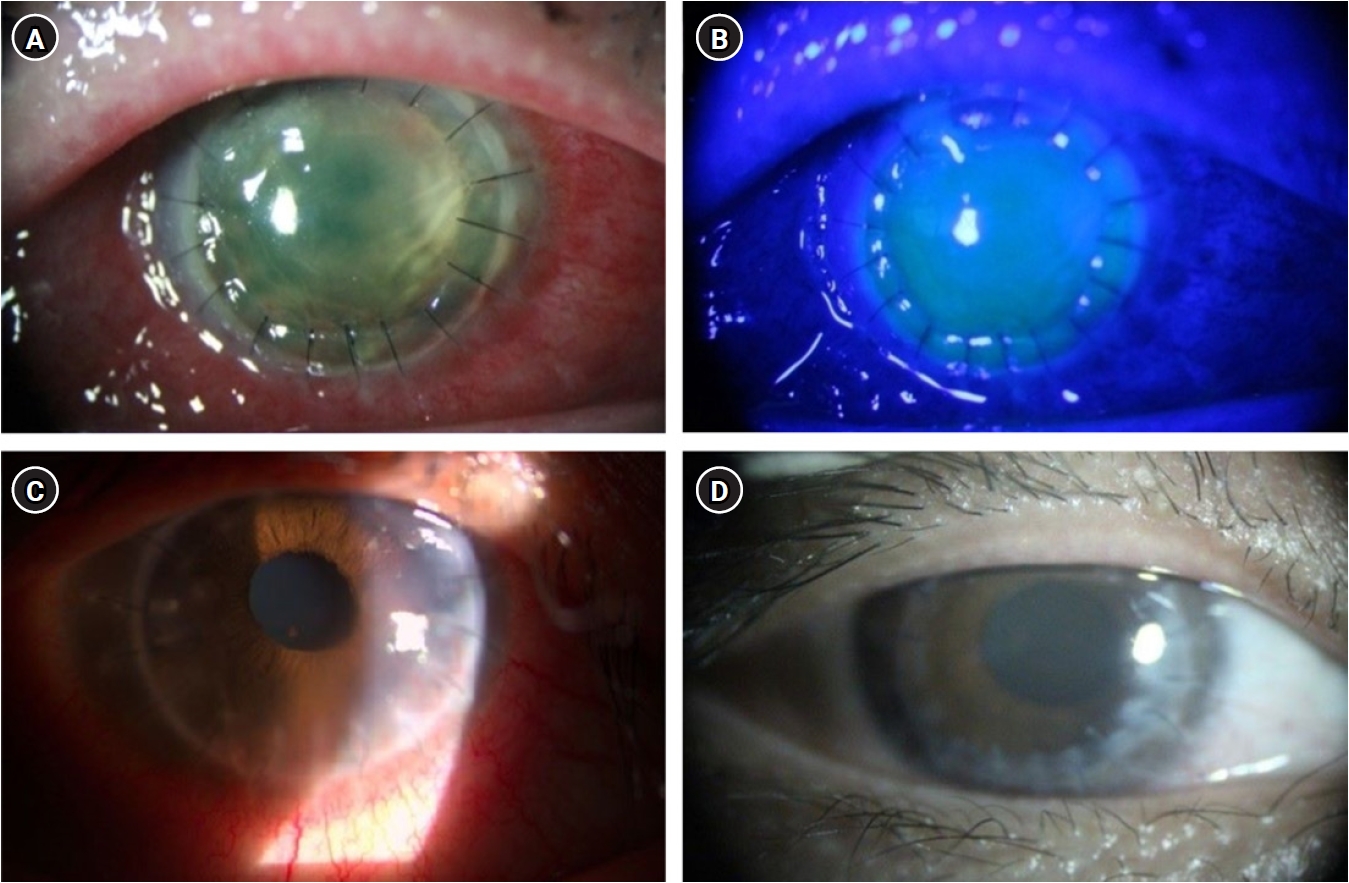
Topical antiamoebic, antibacterial, and ocular hypotensive agents were initiated. However, given the uncontrolled amoebic infection, emergency therapeutic PKP was performed the following day to reduce the substantial infectious burden (Fig. 3A, B). The recipient cornea was trephined to 7.25 mm, and an oversized 8.25 mm donor corneal button was secured with 16 interrupted 10-0 nylon sutures. Intraoperatively, anterior chamber irrigation with moxifloxacin and 1% voriconazole was performed. The crystalline lens was left intact without intraoperative manipulation. Intraoperative corneal cultures were obtained for microbiological evaluation. Histopathological examination of the excised corneal button demonstrated degenerated epithelioid cell aggregates with features suggestive of encysted organisms, consistent with amoebic infection accompanied by fibroinflammatory changes. Special stains, including Gomori methenamine silver and periodic acid-Schiff, were negative for fungal organisms, and immunohistochemical markers were also negative. Microbiological cultures showed no bacterial or fungal growth.
Given the risk of Acanthamoeba keratitis recurrence, topical corticosteroids were deliberately withheld in the immediate postoperative period. Antiamoebic therapy consisted of topical PHMB (0.02%) alone, administered intensively during the first 2 postoperative weeks with dosing adjusted from hourly to every 2 to 4 hours according to the clinical course. Subsequently, the dosing was reduced to four times daily from 3 weeks, tapered to twice daily at 8 weeks, and discontinued approximately 10 weeks postoperatively. Topical moxifloxacin was also administered as prophylactic antimicrobial therapy. As postoperative inflammation increased, systemic corticosteroids were introduced on postoperative day 7 to control inflammation while minimizing the risk of infection recurrence associated with topical steroid use. Immunosuppression was subsequently intensified with systemic corticosteroids and the addition of mycophenolate mofetil. Topical corticosteroids were introduced approximately 1 month postoperatively, once the risk of recurrent Acanthamoeba infection was considered low.
During follow-up, early signs of graft rejection, including corneal neovascularization at the graft–host junction, were observed at 4 weeks postoperatively (Fig. 3C). Immunosuppressive therapy was intensified accordingly. Suture loosening was subsequently managed with corneal re-suturing. Due to recurrent suture loosening with progressive neovascularization, a second re-suturing procedure with subconjunctival dexamethasone and bevacizumab injection was performed, followed by additional bevacizumab injection during follow-up.
At 4 months postoperatively, the graft remained clear without evidence of recurrent infection (Fig. 3D). BCVA improved to 0.2 at approximately 3 weeks postoperatively, and to 0.7 with manifest refraction at the final follow-up, approximately 5 to 6 months postoperatively. IOP normalized following surgery and remained within normal range throughout the postoperative course without further hypotensive therapy.
Acanthamoeba keratitis remains a challenging corneal infection due to its resistance to conventional antimicrobial therapy and its potential for rapid progression. Treatment requires prolonged administration of topical antiamoebic agents, as cyst forms are relatively resistant and may lead to recurrence [6]. Premature tapering of therapy or inappropriate adjunctive treatments, particularly corticosteroid use before complete eradication of Acanthamoeba, may lead to disease exacerbation [7].
In the present case, long-term orthokeratology lens use combined with tap water–based care likely contributed to both disease acquisition and severity [2,8]. The clinical course was further influenced by early treatment modification. Antiamoebic therapy was not maintained continuously, and amniotic membrane transplantation was performed, followed by corticosteroid use prior to confirmed eradication of Acanthamoeba. Given that Acanthamoeba keratitis requires prolonged exposure to antiamoebic agents, early discontinuation of PHMB in combination with corticosteroid use may have contributed to disease progression, resulting in rapid stromal deterioration and extensive corneal involvement.
In cases where infection is uncontrolled, timely surgical intervention becomes critical to preserving ocular integrity. PKP in Acanthamoeba keratitis is generally classified as either optical or therapeutic. Optical PKP is typically performed after complete infection control and is associated with better graft survival and visual outcomes [9]. In contrast, therapeutic PKP is reserved for cases with progressive stromal melting or impending corneal perforation despite maximal medical therapy [10]. The optimal timing of PKP in Acanthamoeba keratitis remains controversial, with increasing emphasis on individualized decision-making [9,11]. Recent studies suggest that earlier surgical intervention may be beneficial in selected cases by limiting irreversible stromal damage [6,12]. The concept of “low-load keratoplasty” in which surgery is performed after partial reduction of the infectious burden, further supports a more flexible, individualized approach to surgical timing [13].
Consistent with these considerations, emergency therapeutic PKP was performed as a globe-preserving intervention in the setting of extensive stromal melting and elevated IOP, findings suggestive of uncontrolled infection. This case illustrates that early therapeutic PKP is appropriate when progressive tissue destruction indicates uncontrolled infection with a high risk of corneal perforation.
Therapeutic PKP in Acanthamoeba keratitis poses a distinct postoperative challenge, as graft survival must be balanced against the risk of persistent or recurrent infection. In routine keratoplasty, early intensive topical corticosteroid therapy is commonly used to reduce the risk of immune-mediated graft rejection [14]. In Acanthamoeba keratitis, corticosteroid use before adequate antiamoebic control has been associated with worse outcomes, and early postoperative corticosteroid exposure may increase the risk of recurrence [7].
In the present case, a staged immunosuppressive approach was adopted. Given that topical corticosteroids act directly on the corneal surface where Acanthamoeba cysts may persist, systemic corticosteroids were introduced first to mitigate graft rejection while minimizing direct interference with local antiamoebic defense. Mycophenolate mofetil was subsequently added to provide additional immunosuppression for graft survival through inhibition of lymphocyte proliferation. Topical corticosteroids were deferred until the risk of persistent infection was considered lower. This stepwise strategy allowed rejection to be addressed while maintaining vigilance for recurrent Acanthamoeba keratitis.
Several limitations warrant consideration in interpreting the findings of this report. As a single case report, the findings may not be generalizable to all clinical presentations of Acanthamoeba keratitis. Furthermore, the relatively short follow-up period of 5 to 6 months limits our ability to draw definitive conclusions regarding longer-term outcomes, including graft survival, visual stability, recurrent infection, and immune rejection. Future prospective studies or larger case series are needed to validate the optimal timing of therapeutic PKP and the long-term efficacy of a staged immunosuppressive approach in the management of severe Acanthamoeba keratitis.
Nevertheless, this case highlights the complex management of severe Acanthamoeba keratitis, in which timely surgical intervention and careful postoperative management are critical to achieving favorable outcomes. The timing of PKP should be individualized, with emergency therapeutic PKP indicated in cases of uncontrolled infection with a high risk of corneal perforation. Even after surgery, outcomes depend on careful balancing of graft rejection against the risk of persistent or recurrent infection, necessitating a cautious and case-specific approach to immunosuppression.

Author contributions

Conceptualization: TIK. Data curation: YC. Formal analysis: JJ. Investigation: YC, JJ. Methodology: YC, JJ. Project administration: TIK. Resources: TIK. Software: JJ. Supervision: TIK. Validation: YC, JJ. Visualization: YC. Writing – original draft: YC, JJ. Writing – review & editing: all authors. Final approval of the manuscript: all authors.

Conflicts of interest

No potential conflict of interest relevant to this article was reported.

Funding

None.

Data availability

Not applicable.

Fig. 1.
In vivo confocal microscopy findings at the time of diagnosis, showing multiple hyperreflective, double-walled cyst-like structures (arrows).
icrs-26-006f1.jpg
Fig. 2.
Preoperative slit-lamp biomicroscopic findings. (A) Corneal epithelial erosion with ring infiltration after discontinuation of antiamoebic therapy at the referring hospital before amniotic membrane transplantation. (B) Progressive diffuse stromal infiltration with hypopyon after amniotic membrane transplantation and adjunctive topical corticosteroid use. (C) Dense corneal infiltration with a large epithelial defect and central corneal thinning at presentation to our clinic. (D) Central corneal epithelial defect with fluorescein staining.
icrs-26-006f2.jpg
Fig. 3.
Postoperative slit-lamp biomicroscopic findings after therapeutic penetrating keratoplasty. (A) Postoperative day 1, showing the corneal graft in place. (B) Incomplete graft epithelialization with fluorescein staining on postoperative day 1. (C) Corneal neovascularization at the inferior graft–host junction at 1 month postoperatively. (D) Stable corneal graft without evidence of recurrent infection at 4 months postoperatively.
icrs-26-006f3.jpg
  • 1. Moore MB, McCulley JP, Newton C, et al. Acanthamoeba keratitis: a growing problem in soft and hard contact lens wearers. Ophthalmology 1987;94:1654‒61. PubMed
  • 2. Wu J, Xie H. Orthokeratology lens-related Acanthamoeba keratitis: case report and analytical review. J Int Med Res 2021;49:3000605211000985.ArticlePubMedPMCPDF
  • 3. Raghavan A, Rammohan R. Acanthamoeba keratitis: a review. Indian J Ophthalmol 2024;72:473‒82. ArticlePubMedPMC
  • 4. Szentmary N, Daas L, Shi L, et al. Acanthamoeba keratitis: clinical signs, differential diagnosis and treatment. J Curr Ophthalmol 2018;31:16‒23. ArticlePubMedPMC
  • 5. Theodore FH, Jakobiec FA, Juechter KB, et al. The diagnostic value of a ring infiltrate in acanthamoebic keratitis. Ophthalmology 1985;92:1471‒9. ArticlePubMed
  • 6. Fu L, Wasielica-Poslednik J, Geerling G, et al. Navigating the challenges of Acanthamoeba keratitis: current trends and future directions. Life (Basel) 2025;15:933.ArticlePubMedPMC
  • 7. Varacalli G, Di Zazzo A, Mori T, et al. Challenges in Acanthamoeba keratitis: a review. J Clin Med 2021;10:942.ArticlePubMedPMC
  • 8. Tseng CH, Fong CF, Chen WL, et al. Overnight orthokeratology-associated microbial keratitis. Cornea 2005;24:778‒82. ArticlePubMed
  • 9. Di Zazzo A, Varacalli G, De Gregorio C, et al. Therapeutic corneal transplantation in Acanthamoeba keratitis: penetrating versus lamellar keratoplasty. Cornea 2022;41:396‒401. ArticlePubMed
  • 10. Dart JK, Saw VP, Kilvington S. Acanthamoeba keratitis: diagnosis and treatment update 2009. Am J Ophthalmol 2009;148:487‒99. ArticlePubMed
  • 11. Laurik KL, Szentmary N, Daas L, et al. Early penetrating keratoplasty à chaud may improve outcome in therapy-resistant Acanthamoeba keratitis. Adv Ther 2019;36:2528‒40. ArticlePubMedPDF
  • 12. Jiang S, Wang L, Zhang Z, et al. Corneal transplantation strategies for Acanthamoeba keratitis: a systematic review and meta-analysis. J Infect Public Health 2026;19:103114.ArticlePubMed
  • 13. Abu Dail Y, Flockerzi E, Munteanu C, et al. Rethinking keratoplasty for patients with Acanthamoeba keratitis: early “Low Load Keratoplasty” in contrast to late optical and therapeutic keratoplasty. Microorganisms 2024;12:1801.ArticlePubMedPMC
  • 14. Azevedo Magalhaes O, Shalaby Bardan A, Zarei-Ghanavati M, Liu C. Literature review and suggested protocol for prevention and treatment of corneal graft rejection. Eye (Lond) 2020;34:442‒50. ArticlePubMedPDF

Figure & Data

References

    Citations

    Citations to this article as recorded by  

      • ePub LinkePub Link
      • Cite
        CITE
        export Copy Download
        Close
        Download Citation
        Download a citation file in RIS format that can be imported by all major citation management software, including EndNote, ProCite, RefWorks, and Reference Manager.

        Format:
        • RIS — For EndNote, ProCite, RefWorks, and most other reference management software
        • BibTeX — For JabRef, BibDesk, and other BibTeX-specific software
        Include:
        • Citation for the content below
        Therapeutic penetrating keratoplasty in rapidly progressive Acanthamoeba keratitis: a case report
        Close
      • XML DownloadXML Download
      Figure
      • 0
      • 1
      • 2
      We recommend
      Therapeutic penetrating keratoplasty in rapidly progressive Acanthamoeba keratitis: a case report
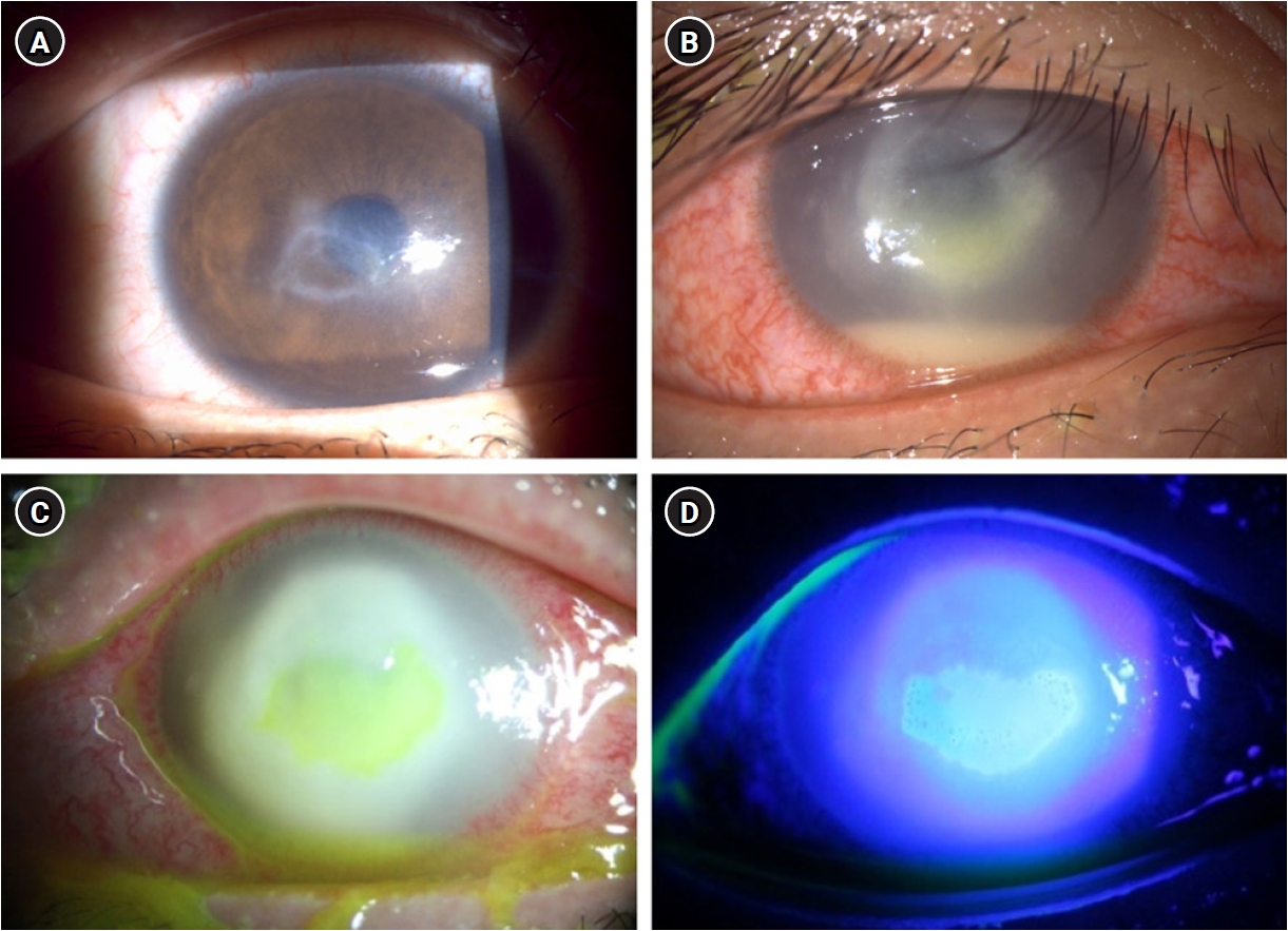
      Image Image Image
      Fig. 1. In vivo confocal microscopy findings at the time of diagnosis, showing multiple hyperreflective, double-walled cyst-like structures (arrows).
      Fig. 2. Preoperative slit-lamp biomicroscopic findings. (A) Corneal epithelial erosion with ring infiltration after discontinuation of antiamoebic therapy at the referring hospital before amniotic membrane transplantation. (B) Progressive diffuse stromal infiltration with hypopyon after amniotic membrane transplantation and adjunctive topical corticosteroid use. (C) Dense corneal infiltration with a large epithelial defect and central corneal thinning at presentation to our clinic. (D) Central corneal epithelial defect with fluorescein staining.
      Fig. 3. Postoperative slit-lamp biomicroscopic findings after therapeutic penetrating keratoplasty. (A) Postoperative day 1, showing the corneal graft in place. (B) Incomplete graft epithelialization with fluorescein staining on postoperative day 1. (C) Corneal neovascularization at the inferior graft–host junction at 1 month postoperatively. (D) Stable corneal graft without evidence of recurrent infection at 4 months postoperatively.
      Therapeutic penetrating keratoplasty in rapidly progressive Acanthamoeba keratitis: a case report
      Insights Cataract Refract Surg : Insights in Cataract and Refractive Surgery
      Close layer
      TOP
