
 KSCRS
KSCRS
Articles
- Page Path
- HOME > Insights Cataract Refract Surg > Ahead-of print articles > Article
-
Original Article
연속초점 인공수정체에서 난시 인공수정체와 윤부이완절개술의 효과 비교 -
이지영, 김은철

-
Comparison of toric intraocular lenses and limbal relaxing incision in extended-depth-of-focus intraocular lens implantation -
Jiyoung Emily Lee, Eun Chul Kim
-
DOI: https://doi.org/10.63375/icrs.26.003
Published online: September 23, 2026
가톨릭대학교 의과대학 부천성모병원 안과학교실
Department of Ophthalmology, Bucheon St. Mary’s Hospital, College of Medicine, The Catholic University of Korea, Seoul, Korea
- Correspondence to: Eun Chul Kim Department of Ophthalmology, Bucheon St. Mary’s Hospital, College of Medicine, The Catholic University of Korea, 327 Sosa-ro, Wonmi-gu, Bucheon 14647, Korea Tel: +82-32-340-2125 E-mail: eunchol@hanmail.net
• Received: March 30, 2026 • Revised: August 11, 2026 • Accepted: August 14, 2026
© 2026 Korean Society of Cataract and Refractive Surgery.
This is an Open Access article distributed under the terms of the Creative Commons Attribution Non-Commercial License (https://creativecommons.org/licenses/by-nc/4.0/) which permits unrestricted non-commercial use, distribution, and reproduction in any medium, provided the original work is properly cited.
- 46 Views
- 0 Download


국문초록
-
목적
- 백내장 수술 시 연속초점 인공수정체에서 난시 인공수정체와 윤부이완절개술의 시력 효과를 비교하였다.
-
방법
- 연속초점 인공수정체를 이용하여 백내장 수술을 받은 환자들 중 난시 인공수정체를 사용하여 수술 시행한 60명의 환자군과 윤부이완절개술을 시행한 60명의 환자군을 대상으로 수술 후 임상 경과를 비교하였다. 각막난시가 1.0 diopters (D)를 초과할 때 난시 인공수정체를 삽입하였고(1군), 각막난시가 1.0 D 이하인 경우 윤부이완절개술을 시행하였다(2군). 각 군에서 수술 전과 수술 3개월 후 각막난시, 굴절난시, 굴절률, 나안시력, 교정시력에 대하여 분석하였다.
-
결과
- 수술 전과 수술 3개월 후의 각막난시와 전체 각막난시를 비교한 결과, 1군에서 통계적으로 유의한 변화는 없었으나(P>0.05), 2군에서는 수술 후(0.60±0.13 D, 0.61±0.28 D)가 수술 전(0.81±0.36 D, 1.25±0.32 D)에 비해 각각 유의하게 감소하였다. 굴절난시는 1군과 2군 모두 수술 전(2.05±0.64 D, 1.18±0.41 D)보다 수술 3개월 후(0.56±0.32 and 0.62±0.26 D)에 유의하게 감소하였으나, 1군의 감소 폭이 2군보다 유의하게 컸다(P<0.05). 수술 3개월 후 굴절난시는 두 군 모두 통계적인 차이는 없었다(P>0.05). 1군과 2군에서 모두 수술 3개월 후 나안시력과 교정시력이 수술 전 나안시력과 교정시력보다 유의하게 개선되었으나(P<0.05), 두 군 간의 차이는 없었다(P>0.05).
-
결론
- 경도의 난시에는 연속초점 인공수정체와 윤부이완절개술 병행이 효과적일 수 있으며, 중등도 이상의 난시에서는 연속초점 난시 인공수정체를 선택하는 것이 환자의 시력 개선에 도움이 된다.
Abstract
-
Purpose
- This study aimed to compare visual and refractive outcomes after toric intraocular lens (IOL) implantation and limbal relaxing incision (LRI) in eyes receiving extended-depth-of-focus (EDOF) IOLs.
-
Methods
- Clinical outcomes were compared between 60 patients who underwent EDOF toric IOL implantation and 60 patients who underwent EDOF IOL implantation combined with LRI. A toric IOL was implanted when corneal astigmatism exceeded 1.0 diopters (D) (group 1), whereas LRI was performed when corneal astigmatism was 1.0 D or less (group 2). Corneal astigmatism, refractive astigmatism, refraction, uncorrected visual acuity, and corrected visual acuity (logMAR) were analyzed in each group before surgery and 3 months postoperatively.
-
Results
- Corneal and total corneal astigmatism did not differ significantly between preoperative and postoperative measurements in group 1 (P>0.05). In group 2, postoperative corneal and total corneal astigmatism (0.60±0.13 and 0.61±0.28 D, respectively) were significantly lower than the corresponding preoperative values (0.81±0.36 and 1.25±0.32 D, respectively; P<0.05). Refractive astigmatism decreased significantly in both group 1 and group 2, from 2.05±0.64 and 1.18±0.41 D preoperatively to 0.56±0.32 and 0.62±0.26 D at 3 months postoperatively, respectively; however, the magnitude of reduction was significantly greater in group 1 than in group 2 (P<0.05). Postoperative refractive astigmatism did not differ significantly between the two groups (P>0.05). In both groups, uncorrected and corrected visual acuity improved significantly after surgery (P<0.05), with no significant between-group difference (P>0.05).
-
Conclusion
- Combining an EDOF IOL with LRI may be effective for mild astigmatism, whereas selecting a toric EDOF IOL may better support visual improvement in patients with moderate to severe astigmatism.
서론
방법
난시 교정 인공수정체군
윤부이완절개술군
결과
고찰
Article Information
Author contributions
Conceptualization: ECK. Data curation: all authors. Formal analysis: all authors. Visualization: Jall authors. Writing – original draft: all authors. Writing – review & editing: ECK. Final approval of the manuscript: all authors.
Conflicts of interest
No potential conflict of interest relevant to this article was reported.
Funding
None.
Data availability
The data that support the findings of this study are available from the corresponding author upon reasonable request.
Fig. 1.Preoperative and postoperative astigmatism in both groups. In group 2, postoperative corneal astigmatism and TCA (0.60±0.13 and 0.61±0.28 D, respectively) were significantly lower than the corresponding preoperative values (0.81±0.36 and 1.25±0.32 D, respectively; P<0.05). RA decreased significantly in both group 1 and group 2, from 2.05±0.64 and 1.18±0.41 D preoperatively to 0.56±0.32 and 0.62±0.26 D postoperatively, respectively; however, the reduction was significantly greater in group 1 than in group 2 (P<0.05). The Wilcoxon signed-rank test was used for within-group comparisons between preoperative and postoperative data, and the Mann-Whitney U test was used for between-group comparisons. D, diopters; KA, keratometric astigmatism; RA, refractive astigmatism; TCA, total corneal astigmatism. a)Extended-depth-of-focus (EDOF) toric intraocular lens (IOL) implantation for corneal astigmatism >1.0 D; b)EDOF IOL implantation with limbal relaxing incision for corneal astigmatism ≤1.0 D.
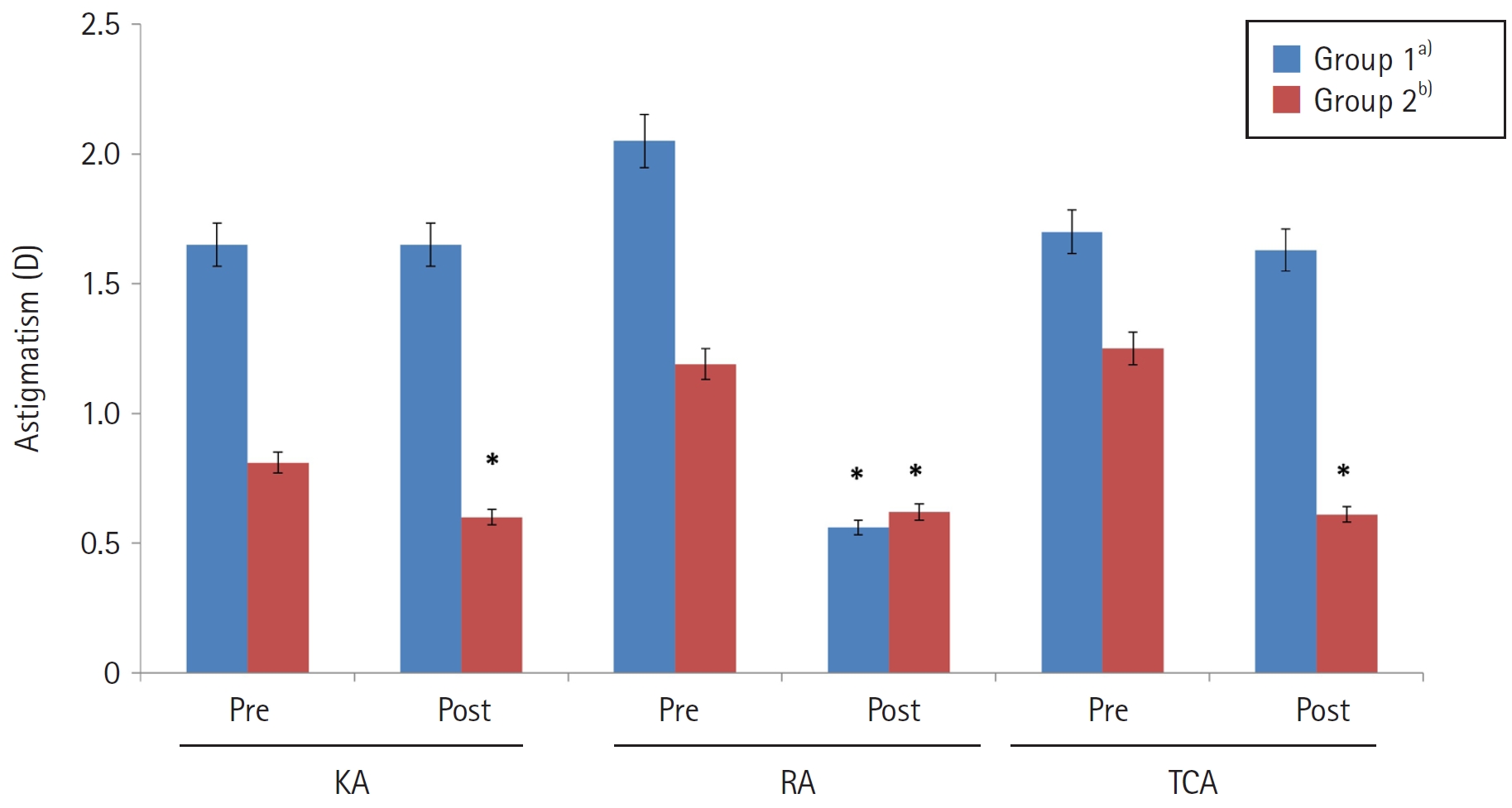
Fig. 2.Preoperative and postoperative visual acuities (logMAR) in both groups. In both groups, postoperative uncorrected and corrected visual acuity improved significantly compared with preoperative values (P<0.05), with no significant difference between groups (P>0.05). The Wilcoxon signed-rank test was used for within-group comparisons between preoperative and postoperative data, and the Mann-Whitney U test was used for between-group comparisons. logMAR, logarithm of the minimum angle of resolution; UCVA, uncorrected visual acuity; BCVA, best-corrected visual acuity; UDVA, uncorrected distance visual acuity; UIVA, uncorrected intermediate visual acuity; CIVA, corrected intermediate visual acuity; UNVA, uncorrected near visual acuity; CNVA, corrected near visual acuity. a)Extended-depth-of-focus (EDOF) toric intraocular lens (IOL) implantation for corneal astigmatism >1.0 D; b)EDOF IOL implantation with limbal relaxing incision for corneal astigmatism ≤1.0 D.
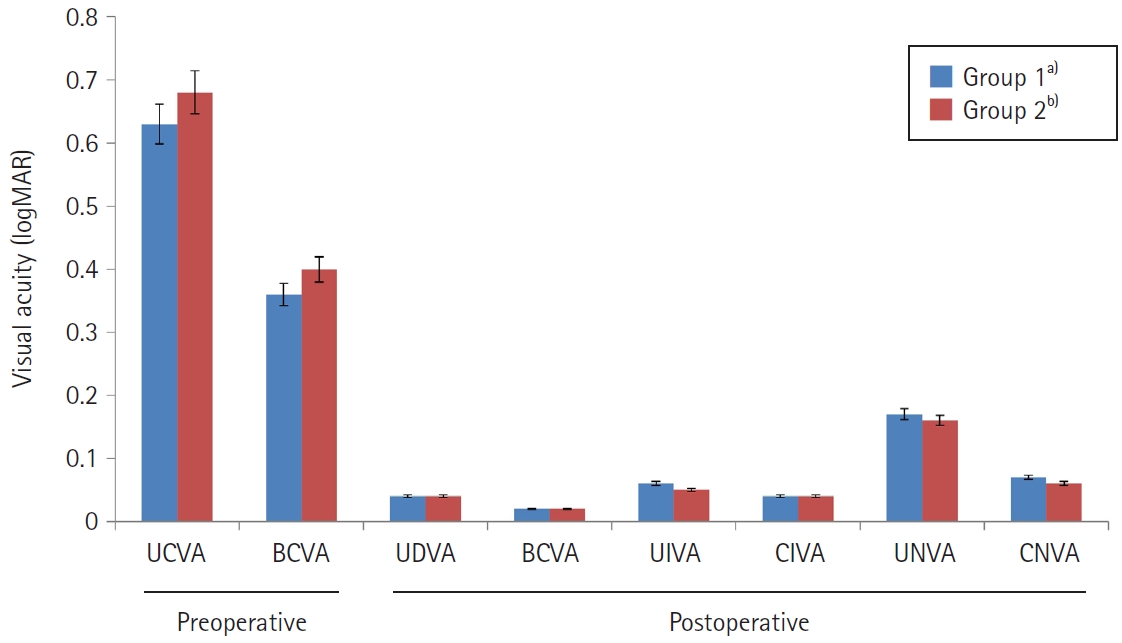
Fig. 3.Mean defocus curves in both groups. Both groups showed peak visual acuity at 0 D defocus (4 m), a relatively smooth profile across the intermediate range (–1.5 to –2.0 D), and a marked decrease in visual acuity in the near range (–3.0 D). logMAR, logarithm of the minimum angle of resolution; D, diopters. a)Extended-depth-of-focus (EDOF) toric intraocular lens (IOL) implantation for corneal astigmatism >1.0 D; b)EDOF IOL implantation with limbal relaxing incision for corneal astigmatism ≤1.0 D.
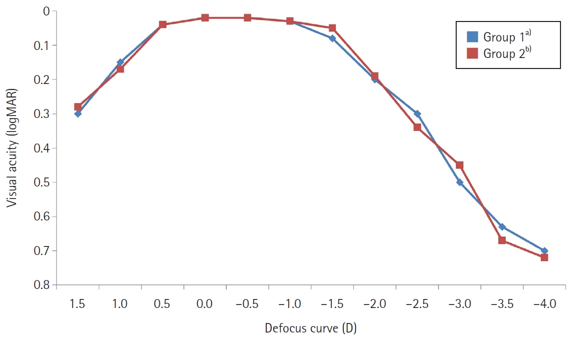
Table 1.Preoperative patient characteristics
| Parameter | Group 1a) | Group 2b) |
|---|---|---|
| Mean age (yr) | 69.5±8.3 | 68.3±9.8 |
| Sex, male:female | 21:39 | 25:35 |
| MRSE (D) | 1.53±2.09 | 1.82±2.36 |
| Keratometric astigmatism (D) | 1.65±0.56* | 0.81±0.36 |
| Refractive astigmatism (D) | 2.05±0.64* | 1.18±0.41 |
| Total corneal astigmatism (D) | 1.70±0.54* | 1.25±0.32 |
| UCVA (logMAR) | 0.63±0.28 | 0.68±0.35 |
| BCVA (logMAR) | 0.41±0.25 | 0.43±0.32 |
| Corneal endothelial cell density (cells/mm2) | 2,636.18±367.52 | 2,594.59±385.29 |
| Total no. of eyes | 60 | 60 |
Values are presented as mean±standard deviation. Preoperative keratometric, refractive, and total corneal astigmatism of group 1 were significantly higher than those of group 2, respectively (*P<0.05). The Mann-Whitney U test was used for between-group comparisons.
MRSE, manifest refraction spherical equivalent; D, diopter; UCVA, uncorrected visual acuity; BCVA, best-corrected visual acuity; logMAR, logarithm of the minimum angle of resolution.
a)Extended-depth-of-focus (EDOF) toric intraocular lens (IOL) implantation for corneal astigmatism >1.0 D;
b)EDOF IOL implantation with limbal relaxing incision for corneal astigmatism ≤1.0 D.
Table 2.Postoperative outcomes at 3 months after surgery
| Parameter | Group 1a) | Group 2b) |
|---|---|---|
| MRSE (D) | –0.18±0.11 | –0.15±0.12 |
| Estimated residual astigmatism (D) | 0.15±0.11 | 0.17±0.12 |
| Postoperative refractive astigmatism (D) | 0.56±0.32 | 0.62±0.26 |
| BCVA (logMAR) | 0.02±0.01 | 0.02±0.01 |
| UDVA (logMAR) | 0.04±0.02 | 0.04±0.02 |
| UIVA (logMAR) | 0.06±0.03 | 0.05±0.02 |
| UNVA (logMAR) | 0.17±0.08 | 0.16±0.07 |
| Corneal endothelial cell loss (cells/mm2) | 265.23±83.26 | 287.59±92.76 |
| Total no. of eyes | 60 | 60 |
Values are presented as mean±standard deviation. No postoperative parameter differed significantly between the two groups (P>0.05). The Mann-Whitney U test was used for between-group comparisons.
MRSE, manifest refraction spherical equivalent; D, diopter; BCVA, best-corrected visual acuity; logMAR, logarithm of the minimum angle of resolution; UDVA, uncorrected distance visual acuity; UIVA, uncorrected intermediate visual acuity; UNVA, uncorrected near visual acuity.
a)Extended-depth-of-focus (EDOF) toric intraocular lens (IOL) implantation for corneal astigmatism >1.0 D;
b)EDOF IOL implantation with limbal relaxing incision for corneal astigmatism ≤1.0 D.
- 1. Hayashi K, Hayashi H, Nakao F, Hayashi F. Influence of astigmatism on multifocal and monofocal intraocular lenses. Am J Ophthalmol 2000;130:477‒82. ArticlePubMed
- 2. Hayashi K, Manabe S, Yoshida M, Hayashi H. Effect of astigmatism on visual acuity in eyes with a diffractive multifocal intraocular lens. J Cataract Refract Surg 2010;36:1323‒9. ArticlePubMed
- 3. Hoffmann PC, Hutz WW. Analysis of biometry and prevalence data for corneal astigmatism in 23,239 eyes. J Cataract Refract Surg 2010;36:1479‒85. ArticlePubMed
- 4. Carvalho MJ, Suzuki SH, Freitas LL, et al. Limbal relaxing incisions to correct corneal astigmatism during phacoemulsification. J Refract Surg 2007;23:499‒504. ArticlePubMed
- 5. Ouchi M, Kinoshita S. Prospective randomized trial of limbal relaxing incisions combined with microincision cataract surgery. J Refract Surg 2010;26:594‒9. ArticlePubMed
- 6. Muftuoglu O, Dao L, Cavanagh HD, et al. Limbal relaxing incisions at the time of apodized diffractive multifocal intraocular lens implantation to reduce astigmatism with or without subsequent laser in situ keratomileusis. J Cataract Refract Surg 2010;36:456‒64. ArticlePubMed
- 7. Kim DH, Wee WR, Lee JH, Kim MK. The short term effects of a single limbal relaxing incision combined with clear corneal incision. Korean J Ophthalmol 2010;24:78‒82. ArticlePubMedPMC
- 8. Muftuoglu O, Prasher P, Chu C, et al. Laser in situ keratomileusis for residual refractive errors after apodized diffractive multifocal intraocular lens implantation. J Cataract Refract Surg 2009;35:1063‒71. ArticlePubMed
- 9. Gangwani V, Hirnschall N, Findl O, Maurino V. Multifocal toric intraocular lenses versus multifocal intraocular lenses combined with peripheral corneal relaxing incisions to correct moderate astigmatism. J Cataract Refract Surg 2014;40:1625‒32. ArticlePubMed
- 10. Vinciguerra P, Holladay JT, Pagano L, et al. Comparison of visual performance and satisfaction with a bilateral emmetropic vs a bilateral mild myopic target using a spherical monofocal intraocular lens. J Cataract Refract Surg 2020;46:839‒43. ArticlePubMed
- 11. Cochener B, Lafuma A, Khoshnood B, et al. Comparison of outcomes with multifocal intraocular lenses: a meta-analysis. Clin Ophthalmol 2011;5:45‒56. ArticlePubMedPMC
- 12. Alarcon A, Canovas C, Koopman B, et al. Enhancing the intermediate vision of monofocal intraocular lenses using a higher order aspheric optic. J Refract Surg 2020;36:520‒7. ArticlePubMed
- 13. Fernandez J, Rocha-de-Lossada C, Zamorano-Martin F, et al. Positioning of enhanced monofocal intraocular lenses between conventional monofocal and extended depth of focus lenses: a scoping review. BMC Ophthalmol 2023;23:101.ArticlePubMedPMC
- 14. Garzon N, Poyales F, de Zarate BO, et al. Evaluation of rotation and visual outcomes after implantation of monofocal and multifocal toric intraocular lenses. J Refract Surg 2015;31:90‒7. ArticlePubMed
- 15. Ouchi M, Kinoshita S. AcrySof IQ toric IOL implantation combined with limbal relaxing incision during cataract surgery for eyes with astigmatism >2.50 D. J Refract Surg 2011;27:643‒7. ArticlePubMed
- 16. Ouchi M. High-cylinder toric intraocular lens implantation versus combined surgery of low-cylinder intraocular lens implantation and limbal relaxing incision for high-astigmatism eyes. Clin Ophthalmol 2014;8:661‒7. ArticlePubMedPMC
- 17. Na KS, An D, Kim HS, Kim EC. Relation between change of effective lens position and toric IOL rotation after toric IOL implantation. Can J Ophthalmol 2025;60:8‒14. ArticlePubMed
- 18. Nichamin LD. Management of astigmatism in conjunction with clear corneal phaco surgery. In: Gills JP, editor. A complete surgical guide for correcting astigmatism: an ophthalmic manifesto. Slack Inc.; 2003. p. 41‒7.
- 19. Kim EC. Efficacy of extended depth of focus, enhanced monofocal, and monofocal intraocular lenses in patients with retinal disease. Insights Cataract Refract Surg 2025;10:52‒60. ArticlePDF
References
Figure & Data
References
Citations
Citations to this article as recorded by 
 ePub Link
 Cite
Cite XML Download
Comparison of toric intraocular lenses and limbal relaxing incision in extended-depth-of-focus intraocular lens implantation
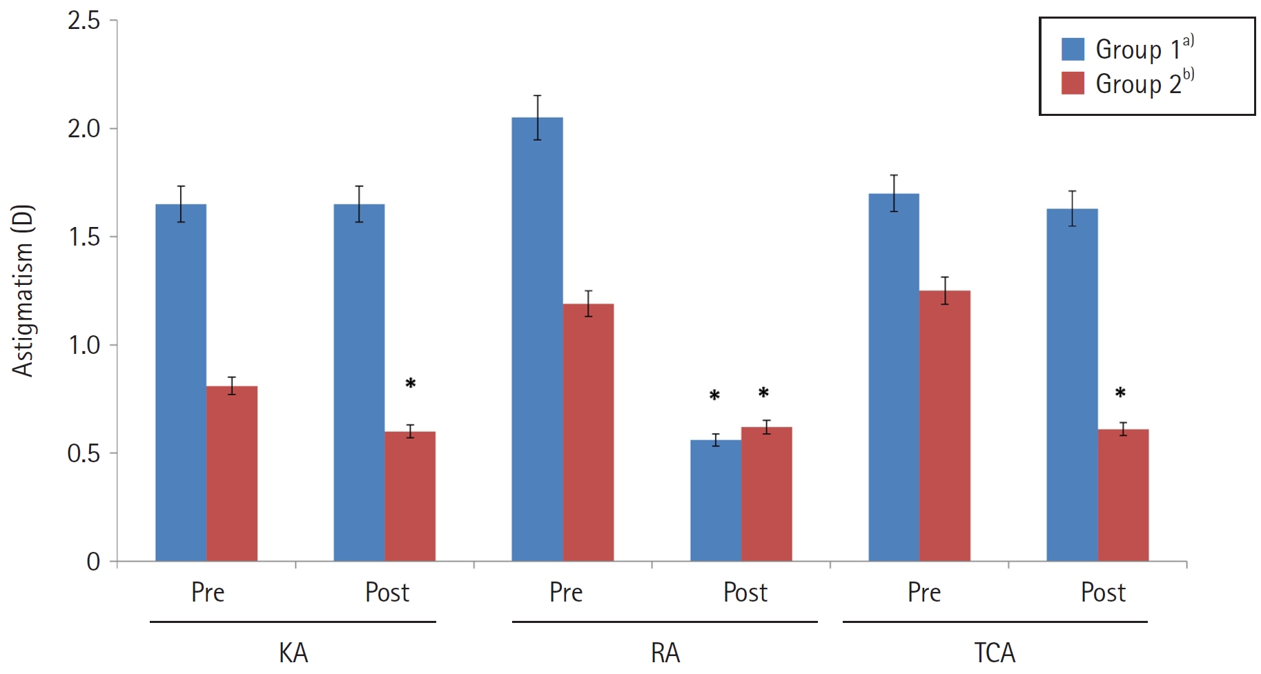
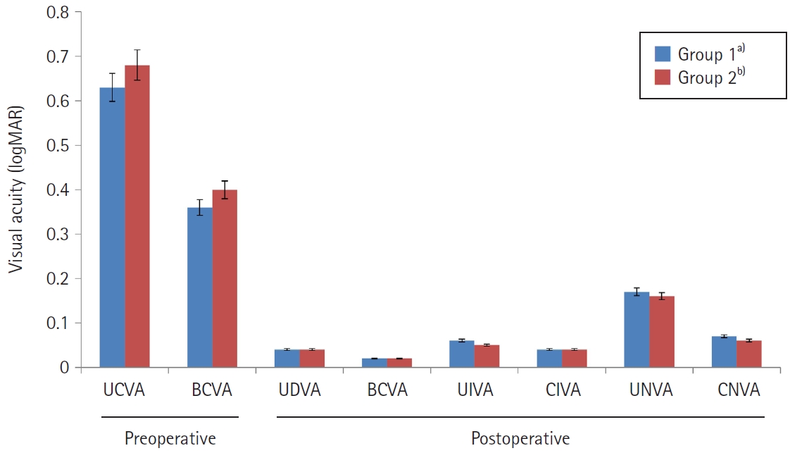
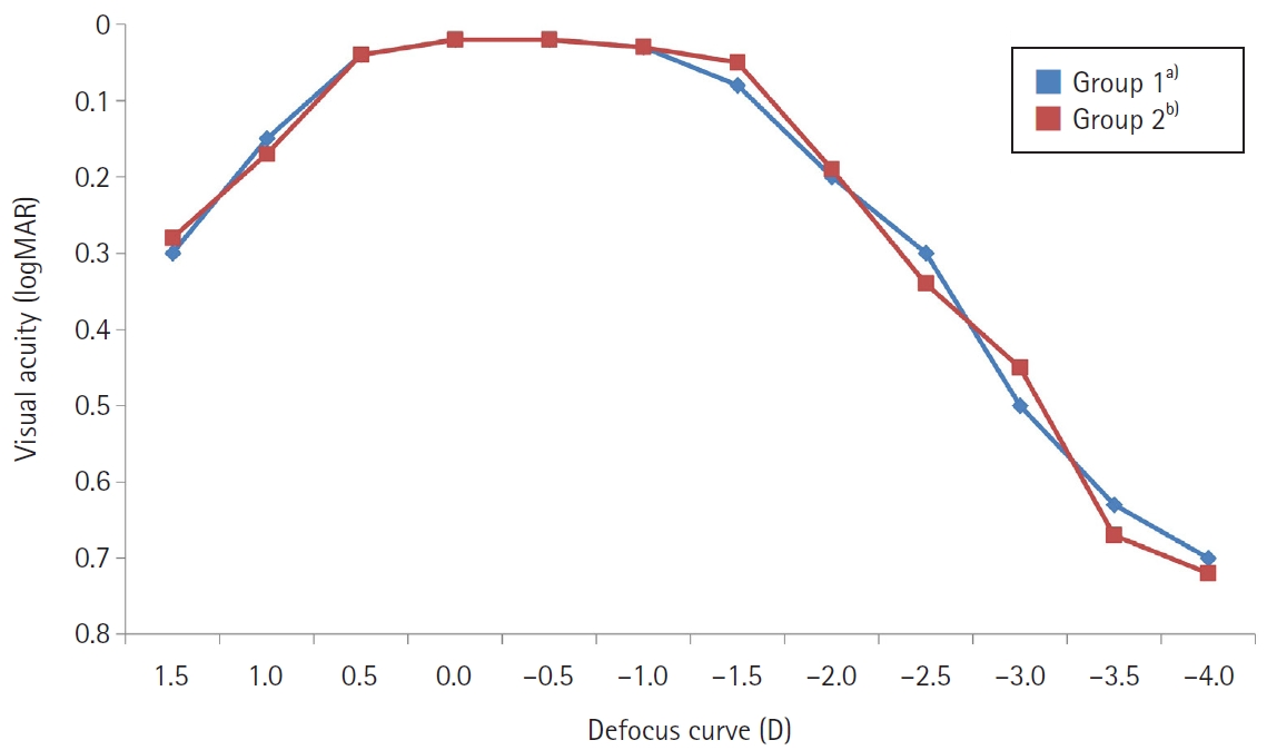
Fig. 1. Preoperative and postoperative astigmatism in both groups. In group 2, postoperative corneal astigmatism and TCA (0.60±0.13 and 0.61±0.28 D, respectively) were significantly lower than the corresponding preoperative values (0.81±0.36 and 1.25±0.32 D, respectively; P<0.05). RA decreased significantly in both group 1 and group 2, from 2.05±0.64 and 1.18±0.41 D preoperatively to 0.56±0.32 and 0.62±0.26 D postoperatively, respectively; however, the reduction was significantly greater in group 1 than in group 2 (P<0.05). The Wilcoxon signed-rank test was used for within-group comparisons between preoperative and postoperative data, and the Mann-Whitney U test was used for between-group comparisons. D, diopters; KA, keratometric astigmatism; RA, refractive astigmatism; TCA, total corneal astigmatism. a)Extended-depth-of-focus (EDOF) toric intraocular lens (IOL) implantation for corneal astigmatism >1.0 D; b)EDOF IOL implantation with limbal relaxing incision for corneal astigmatism ≤1.0 D.
Fig. 2. Preoperative and postoperative visual acuities (logMAR) in both groups. In both groups, postoperative uncorrected and corrected visual acuity improved significantly compared with preoperative values (P<0.05), with no significant difference between groups (P>0.05). The Wilcoxon signed-rank test was used for within-group comparisons between preoperative and postoperative data, and the Mann-Whitney U test was used for between-group comparisons. logMAR, logarithm of the minimum angle of resolution; UCVA, uncorrected visual acuity; BCVA, best-corrected visual acuity; UDVA, uncorrected distance visual acuity; UIVA, uncorrected intermediate visual acuity; CIVA, corrected intermediate visual acuity; UNVA, uncorrected near visual acuity; CNVA, corrected near visual acuity. a)Extended-depth-of-focus (EDOF) toric intraocular lens (IOL) implantation for corneal astigmatism >1.0 D; b)EDOF IOL implantation with limbal relaxing incision for corneal astigmatism ≤1.0 D.
Fig. 3. Mean defocus curves in both groups. Both groups showed peak visual acuity at 0 D defocus (4 m), a relatively smooth profile across the intermediate range (–1.5 to –2.0 D), and a marked decrease in visual acuity in the near range (–3.0 D). logMAR, logarithm of the minimum angle of resolution; D, diopters. a)Extended-depth-of-focus (EDOF) toric intraocular lens (IOL) implantation for corneal astigmatism >1.0 D; b)EDOF IOL implantation with limbal relaxing incision for corneal astigmatism ≤1.0 D.
Fig. 1.
Fig. 2.
Fig. 3.
Comparison of toric intraocular lenses and limbal relaxing incision in extended-depth-of-focus intraocular lens implantation
| Parameter | Group 1 |
Group 2 |
|---|---|---|
| Mean age (yr) | 69.5±8.3 | 68.3±9.8 |
| Sex, male:female | 21:39 | 25:35 |
| MRSE (D) | 1.53±2.09 | 1.82±2.36 |
| Keratometric astigmatism (D) | 1.65±0.56 |
0.81±0.36 |
| Refractive astigmatism (D) | 2.05±0.64 |
1.18±0.41 |
| Total corneal astigmatism (D) | 1.70±0.54 |
1.25±0.32 |
| UCVA (logMAR) | 0.63±0.28 | 0.68±0.35 |
| BCVA (logMAR) | 0.41±0.25 | 0.43±0.32 |
| Corneal endothelial cell density (cells/mm2) | 2,636.18±367.52 | 2,594.59±385.29 |
| Total no. of eyes | 60 | 60 |
| Parameter | Group 1 |
Group 2 |
|---|---|---|
| MRSE (D) | –0.18±0.11 | –0.15±0.12 |
| Estimated residual astigmatism (D) | 0.15±0.11 | 0.17±0.12 |
| Postoperative refractive astigmatism (D) | 0.56±0.32 | 0.62±0.26 |
| BCVA (logMAR) | 0.02±0.01 | 0.02±0.01 |
| UDVA (logMAR) | 0.04±0.02 | 0.04±0.02 |
| UIVA (logMAR) | 0.06±0.03 | 0.05±0.02 |
| UNVA (logMAR) | 0.17±0.08 | 0.16±0.07 |
| Corneal endothelial cell loss (cells/mm2) | 265.23±83.26 | 287.59±92.76 |
| Total no. of eyes | 60 | 60 |
Table 1. Preoperative patient characteristics
Values are presented as mean±standard deviation. Preoperative keratometric, refractive, and total corneal astigmatism of group 1 were significantly higher than those of group 2, respectively (*P<0.05). The Mann-Whitney U test was used for between-group comparisons. MRSE, manifest refraction spherical equivalent; D, diopter; UCVA, uncorrected visual acuity; BCVA, best-corrected visual acuity; logMAR, logarithm of the minimum angle of resolution. Extended-depth-of-focus (EDOF) toric intraocular lens (IOL) implantation for corneal astigmatism >1.0 D; EDOF IOL implantation with limbal relaxing incision for corneal astigmatism ≤1.0 D.
Table 2. Postoperative outcomes at 3 months after surgery
Values are presented as mean±standard deviation. No postoperative parameter differed significantly between the two groups (P>0.05). The Mann-Whitney U test was used for between-group comparisons. MRSE, manifest refraction spherical equivalent; D, diopter; BCVA, best-corrected visual acuity; logMAR, logarithm of the minimum angle of resolution; UDVA, uncorrected distance visual acuity; UIVA, uncorrected intermediate visual acuity; UNVA, uncorrected near visual acuity. Extended-depth-of-focus (EDOF) toric intraocular lens (IOL) implantation for corneal astigmatism >1.0 D; EDOF IOL implantation with limbal relaxing incision for corneal astigmatism ≤1.0 D.


