
Previous issues
- Page Path
- HOME > Browse articles > Previous issues
- Volume 10(3); October 2025
-


Review Article
- Phacoemulsification in patients with diabetes: from preoperative evaluation to postoperative management
- Yeoun Sook Chun
- Insights Cataract Refract Surg 2025;10(3):65-75. Published online October 31, 2025
- DOI: https://doi.org/10.63375/icrs.25.012
-
 Abstract
 PDF
PDF ePub
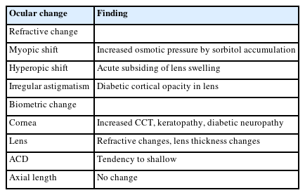
ePub - Diabetes mellitus is one of the most common chronic diseases worldwide and is a leading cause of blindness in patients over the age of 50 years. Patients with diabetes have an elevated risk of developing cataracts compared to individuals without diabetes; furthermore, cataracts also tend to progress more rapidly in this population, leading to the need for surgery at a younger age. This review aims to summarize the key considerations in the management of cataract surgery in patients with diabetes, from preoperative evaluation to postoperative care. Patients with diabetes often present with unstable refractive status, dry eye disease, corneal epithelial defects, and recurrent corneal erosions. They also tend to have reduced corneal endothelial cell density and small pupils, both of which increase the risk of intraoperative complications. Postoperatively, these patients are at risk of developing pseudophakic cystoid macular edema, posterior capsular opacification, endophthalmitis, progression of diabetic retinopathy, and neovascular glaucoma. Patients with long-standing or poorly controlled diabetes face a higher likelihood of postoperative complications, highlighting the importance of regular ophthalmic follow-up examinations. Furthermore, adjunctive treatments such as timely intravitreal injections of anti-vascular endothelial growth factor agents may reduce the risk of vision-threatening complications following cataract surgery.
- 1,252 View
- 14 Download

Original Articles
- Results of multifocal intraocular lens implantation in patients who underwent corneal refractive surgery
- Eun Chul Kim
- Insights Cataract Refract Surg 2025;10(3):76-82. Published online October 31, 2025
- DOI: https://doi.org/10.63375/icrs.25.011
-
Abstract
PDFePub
- Purpose
The aim of this study was to evaluate the clinical results of multifocal intraocular lenses in patients who underwent corneal refractive surgery.
Methods
Thirty eyes (16 patients; Synergy: ZFR00V) were retrospectively enrolled. Uncorrected and corrected near visual acuity (UNVA, CNVA), intermediate visual acuity (UIVA, CIVA), and distant visual acuity (UDVA, CDVA), manifest refraction spherical equivalent (MRSE), and satisfaction score were assessed before and after surgery.
Results
The postoperative UDVA, UIVA, UNVA, and MRSE of the three groups exhibited improvements compared to the preoperative data (P<0.05). The error between the postoperative refraction and the intraocular lens calculation was smaller with the Barrett True K formula than with the Haigis-L formula (P<0.05). The defocus curve at 0 diopter (D) increased, from –1 to –1.5 D, and from –2.5 to –4.0 D, indicating improved vision at distant, intermediate, and near distances. Distance satisfaction (1.47±0.63), near satisfaction (1.25±0.71), and overall satisfaction (1.36±0.42) were good, but light scattering and halo satisfaction (1.97±0.85) yielded a poor result.
Conclusion
In patients with cataracts who underwent corneal refractive surgery, multifocal intraocular lens implantation resulted in excellent uncorrected visual acuity at distant, intermediate, and near distances. However, careful consideration should be given to patient selection due to the incidence of side effects such as glare and halos.
- 908 View
- 14 Download
- Clinical results of combined Descemet membrane keratoplasty and cataract operation (triple Descemet membrane keratoplasty) from imported donor corneas: a retrospective study
- Hyung Keun Lee, Sung Soo Kang, Jin Suk Chun, So Young Kim, Dong Ihll Lee
- Insights Cataract Refract Surg 2025;10(3):83-90. Published online October 31, 2025
- DOI: https://doi.org/10.63375/icrs.25.014
-
Abstract
PDFePub
- Purpose
This study reports the clinical outcomes, after triple Descemet membrane endothelial keratoplasty (DMEK) performed using imported corneas.
Methods
A retrospective study was conducted on 30 eyes of 26 patients who underwent Descemet's membrane keratoplasty concurrently with cataract surgery, referred to as triple DMEK, from January 2023 to June 2025. After routine preoperative examinations for keratoplasty as well as cataract surgery, uneventful DMEK surgery was performed concurrently with cataract surgery. All patients visited the clinic at 1, 3, 6, and 12 months after surgery to observe changes, including uncorrected and best spectacle corrected visual acuity, refractive error, corneal thickness, and endothelial cell density.
Results
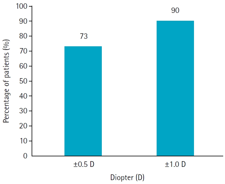
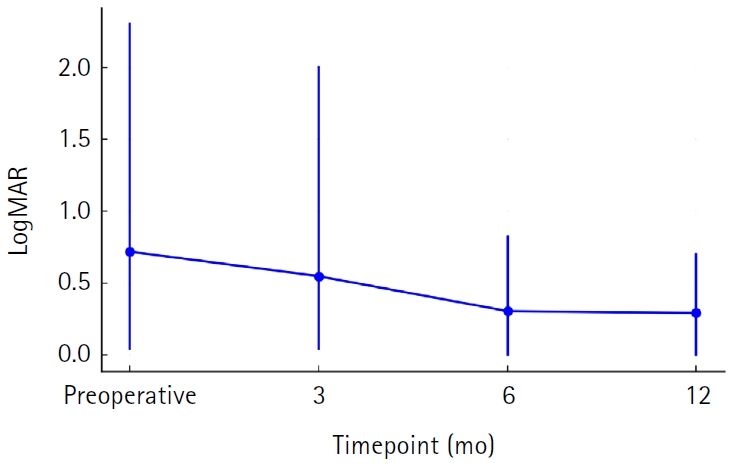
Preoperatively, 12 of the 30 eyes had Fuchs corneal endothelial dystrophy and 12 had endothelial failure following phakic intraocular lens implantation. The average observation period for the subjects was 437±263 days. After triple DMEK surgery, the patients' uncorrected visual acuity gradually improved from 0.73±0.6 (logMAR) before surgery to 0.65±0.54 at 3 months and 0.29±0.26 at 1 year (P<0.001). The change in corneal thickness was 565.7±70.0 μm before surgery, 535.2±44.2 μm at 3 months after surgery, 549.7±73.5 μm at 6 months, and 535.82±49.0 μm at 12 months, but no statistical significance was found compared to before surgery at any time point (P>0.05). The endothelial cell density was 798±363 cells/mm2 before surgery, 1,479±475 cells/mm2 at 3 months after surgery, 1,456±456 cells/mm2 at 6 months, 1,332±346 cells/mm2 at 12 months, and 1,399±519 cells/mm2 at the last visit (P<0.001).
Conclusion
Triple DMEK surgery, which is performed for various corneal diseases, is relatively safe. No significant endothelial damage, refractive changes, or visual acuity abnormalities were observed up to one year after surgery. Future prospective studies including a larger number of participants are warranted to evaluate the safety and clinical outcomes of triple DMEK using imported corneas.
- 848 View
- 7 Download
- The effect of 2% rebamipide ophthalmic solution on early dry eye after SMILE surgery: a retrospective study
- Jin Hyoung Kim, Mu Yan Kim, Young A Kwon, Sung Wook Park, Sung Won Byun, Seong Woo Lee, Sung Hyup Lim
- Insights Cataract Refract Surg 2025;10(3):91-102. Published online October 31, 2025
- DOI: https://doi.org/10.63375/icrs.25.013
-
Abstract
PDFePub
- Purpose
The aim of this study was to investigate the effect of rebamipide 2% ophthalmic solution on early dry eye following small incision lenticule extraction (SMILE) surgery by analyzing dry eye indicators before and after the procedure.
Methods
In this retrospective study, an initial sample of 372 SMILE surgery patients were divided into a rebamipide group (artificial tears and 2% rebamipide) and a control group (artificial tears only). Changes in dry eye indicators, including the Ocular Surface Disease Index (OSDI), corneal fluorescein staining (CFS) score, tear meniscus height (TMH), tear break-up time (TBUT), and dry eye classification, were analyzed at 2 and 4 weeks postoperatively in comparison with the preoperative baseline.
Results
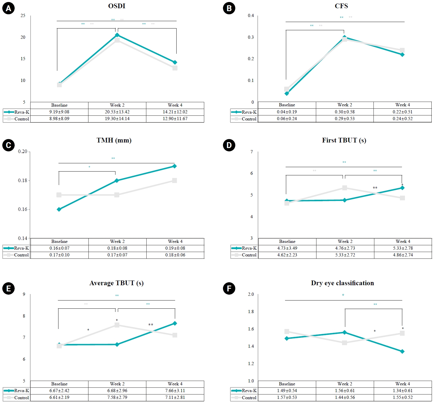
In total, 250 patients (250 eyes) were selected: 135 in the rebamipide group and 115 in the control group. Preoperative characteristics such as gender, age, spherical equivalent refraction, ablation depth, and optical zone size showed no significant differences between the two groups. Both groups demonstrated a significant increase in OSDI and CFS scores at 2 weeks postoperatively, followed by a decrease at 4 weeks, with no significant differences between groups. TMH increased significantly in the rebamipide group at 2 and 4 weeks (P=0.043, P=0.004), but showed no significant change in control group or intergroup difference. No significant difference was found in the first TBUT between the two groups at any time point, but the average TBUT significantly and rapidly increased from 2 to 4 weeks postoperatively in the rebamipide group (P=0.001). The dry eye classification was significantly lower in the rebamipide group at 4 weeks postoperatively (P=0.014).
Conclusion
The use of rebamipide 2% ophthalmic solution immediately after SMILE surgery is expected to be helpful in treating early postoperative dry eye, as it increased TMH and TBUT starting from 2 weeks postoperatively.
- 1,104 View
- 10 Download
 First
First Prev
Prev

