
Skip Navigation
Skip to contents

Insights Cataract Refract Surg : Insights in Cataract and Refractive Surgery

OPEN ACCESS

Articles

Page Path
HOME > Insights Cataract Refract Surg > Volume 11(2); 2026 > Article
Original Article
Effects of extract of Parthenocissus tricuspidata living on pine in a nonclinical model of dry eye disease
Hyeyoon Goo1,2, Chung-Hun Oh3, Kyong Jin Cho2orcid
Insights in Cataract and Refractive Surgery 2026;11(2):56-68.
DOI: https://doi.org/10.63375/icrs.26.005
Published online: June 30, 2026

1Department of Medical Laser, Graduate School, Dankook University College of Medicine, Cheonan, Korea

2Department of Ophthalmology, Dankook University Hospital, Dankook University College of Medicine, Cheonan, Korea

3Department of Oral Physiology, Dankook University College of Medicine, Cheonan, Korea

Correspondence to: Kyong Jin Cho Department of Ophthalmology, Dankook University College of Medicine, 119 Dandae-ro, Dongnam-gu, Cheonan 31116, Korea E-mail: perfectcure@dankook.ac.kr
• Received: May 1, 2026   • Revised: May 28, 2026   • Accepted: May 28, 2026

© 2026 Korean Society of Cataract and Refractive Surgery.

This is an Open Access article distributed under the terms of the Creative Commons Attribution Non-Commercial License (https://creativecommons.org/licenses/by-nc/4.0/) which permits unrestricted non-commercial use, distribution, and reproduction in any medium, provided the original work is properly cited.

  • 233 Views
  • 11 Download
prev
  • Purpose
    Dry eye disease (DED) is a multifactorial ocular surface disorder characterized by tear film instability, hyperosmolarity, and inflammation. Oxidative stress plays an important role in DED pathogenesis by exacerbating ocular surface damage. Parthenocissus tricuspidata growing on pine (PT) has been reported to have antioxidant and anti-inflammatory properties.
  • Methods
    Oxidative stress was induced in human conjunctival epithelial cells (Wong-Kilbourne derivative of Chang conjunctival [WKD] cells) using H2O2, and the antioxidant and protective effects of PT were evaluated. The anti-inflammatory and therapeutic effects of PT were also investigated in a mouse model of DED.
  • Results
    In WKD cells, PT treatment reduced H2O2-induced apoptosis, reactive oxygen species production, and phosphorylation of mitogen-activated protein kinase signaling proteins. Antioxidant enzyme activity, including superoxide dismutase and catalase, increased, whereas malondialdehyde and interleukin-6 levels decreased, indicating reduced oxidative stress and inflammation. In vivo, PT eye drops significantly improved clinical signs of DED, including tear volume and tear film break-up time. Histological analysis and cytokine assays showed reduced expression of pro-inflammatory markers in corneal and conjunctival tissues.
  • Conclusion
    PT extract exerts therapeutic antioxidant and anti-inflammatory effects, highlighting its potential as a treatment for DED.
Dry eye disease (DED) occurs due to defects in eyes and the tear film and is prevalent in dry environments. It is frequently exposed to oxidative stress when exposed to atmospheric oxygen and sunlight, including ultraviolet radiation [1,2]. This oxidative stress induces protein expression [3] and is associated with various eye disease-related and cytokine-related inflammation. Prolonged dryness can lead to corneal surface irregularities, resulting in reduced vision and an increased risk of corneal ulcers [4].
Numerous treatments exist for DED. In mild cases, commonly used artificial tears provide symptomatic relief, albeit temporarily [5]. Another treatment method involves the administration of autologous serum drops, which contain high concentrations of tear constituents, stabilizing the tear film and improving the diseased eye surface. However, frequent blood collection poses a drawback of this treatment [6]. Meanwhile, although corticosteroids have been shown to be effective in clinical studies of corneal epithelial disease, long-term use is associated with toxicity, increased intraocular pressure, and cataracts [7].
Despite the numerous treatments developed for DED, precise therapies remain elusive. Inhibiting oxidative stress is crucial to protect the ocular surface in dry environments [2]. Moreover, considering that reactive oxygen species (ROS) are involved in the inflammatory reaction and antioxidants are effective in treating inflammatory diseases, antioxidants may indirectly inhibit the inflammatory reaction in DED [8,9]. ROS are implicated in various diseases; consequently, research on antioxidants focuses on ROS regulation, with studies identifying natural extracts possessing antioxidant and anti-inflammatory properties [10,11]. Natural extracts have fewer side effects than synthetic extracts and exhibit diverse physiological activities. Therefore, numerous studies have explored their roles as functional materials [11].
Parthenocissus tricuspidata (family Vitaceae) is a vine plant characterized by branched tendrils that attach to surrounding structures [12-14]. Extracts of P. tricuspidata have been reported to possess analgesic, antidiabetic, antioxidant, and anticancer properties [9]. These compounds have been reported to scavenge ROS and suppress inflammatory cytokine expression by regulating oxidative stress-related signaling pathways [10,11]. Because oxidative stress and inflammation are key mechanisms involved in the pathogenesis of DED, natural compounds with antioxidant and anti-inflammatory properties may exert protective effects on the ocular surface under dry conditions [8,9]. Consistently, previous studies have suggested that plant-derived antioxidants can alleviate ocular surface inflammation by modulating ROS-mediated signaling pathways. Although P. tricuspidata living on pine (PT) has shown antioxidant and anti-inflammatory effects at the cellular level, studies evaluating its effects in ocular surface disease models remain limited. Therefore, we investigated the antioxidant and anti-inflammatory effects of PT in in vitro and in vivo models of DED, a major contributor to ocular surface inflammation.
P. tricuspidata extraction and powder manufacturing
P. tricuspidata stems were purchased from Natural Herbs and stored at room temperature. The extraction process proceeded as follows: Briefly, P. tricuspidata and 75% ethanol solvent (diluted in distilled water) were added to an extractor. Following extraction at 80 to 85 ℃ for 8 hours, the extract was sterilized at 65 to 80 ℃ for 30 minutes. The resulting filtrate was concentrated under reduced pressure at a low temperature (65–70 ℃). Subsequently, the concentrate was dried at a high temperature using a dryer, and the resultant powder was dissolved in dimethyl sulfoxide (DMSO; Daejung) and sterile-filtered (Sigma-Aldrich) for experimental use. Before treating cells, the extract was passed through a syringe filter (0.22 μm, Waters Corp.) to remove any remaining impurities. Hereafter, the 75% ethanol extract of P. tricuspidata living on pine is referred to as PT.
The 2,2-diphenyl-1-picrylhydrazyl assay
The 2,2-diphenyl-1-picrylhydrazyl (DPPH) assay was conducted to assess the short-term antioxidant activity of PT; methanol served as the blank. To each test tube, 750 μL of an analytical PT solution, 750 μL of Trolox solution (Sigma-Aldrich), and 300 μL of 0.3 mM DPPH (Sigma-Aldrich) were added. The mixture was stored at room temperature in the dark for 30 minutes, after which the absorbance was measured at 517 nm using a spectrophotometer (Multiskan GO) (Thermo Fisher Scientific). The absorbance resulting from the addition of the analytical PT or control Trolox solution (AS) was normalized against the absorbance of the methanol blank (AM). The radical scavenging activity of DPPH for the analytical PT at each concentration was measured four times. The percent inhibition (%) was calculated using the following equation.
Percent inhibition (%)={1(ASAM)}×100
Cell culture
Wong-Kilbourne derivative of Chang conjunctival cells (WKD cells) (CCL-20, ATCC) were cultured in medium 199 (1×) (Gibco, Thermo Fisher Scientific), supplemented with 1% penicillin-streptomycin (Gibco) and 5% fetal bovine serum (Gibco) at 37 ℃ in a 5% CO2 humid environment. The culture medium was changed every 2 to 3 days. Cells were sub-cultured when the population reached 70% to 80% confluence using 0.25% trypsin-ethylenediaminetetraacetic acid (Corning). Cells between passages 4 and 12 were used for all experiments.
Cell viability assay
Cell viability was assessed using the 3-(4,5-dimethylthiazol-2-yl)-2,5-diphenyltetrazolium bromide (MTT) assay. WKD cells were seeded in 96-well culture plates at a density of 8×10³ cells/well. Cells were pretreated with PT (50, 100, or 500 μg/mL) for 1 hour and then exposed to 70 μM H2O2 for 24 hours. After treatment for 24 hours MTT (Sigma-Aldrich) solution was added to each well. Subsequently, the medium in each well was replaced with DMSO to dissolve the formazan crystals formed, and the absorbance was measured at 575 nm using a spectrophotometer.
Measurement of ROS
Cellular ROS was quantified using the oxidation-sensitive fluorescent dye 2′,7′-dichlorodihydrofluorescein diacetate (DCFH-DA) (Abcam). WKD cells were seeded in black 96-well plates at a density of 8×10³ cells/well for 24 hours and treated with PT for 1 hour. Subsequently, the cell medium in each well was replaced with 20 μΜ DCFH-DA, and the plate was incubated for 30 minutes. After, 70 μΜ H2O2 was added to each well. The formation of fluorescent dichlorofluorescein, resulting from the oxidation of DCFH, was measured at an excitation wavelength of 480 nm and emission wavelength of 520 nm using a fluorescence microplate reader (SpectraMax M2e, Molecular Devices) equipped with the SoftMax Pro software ver. 5 (Molecular Devices).
Protein extraction and Western blotting
Cells were cultured in 6-well plates for 24 hours, followed by pretreatment with PT for 1 hour before exposure to 70 μM H2O2. The following day, the cells were detached by scraping and centrifuged. After centrifugation, the supernatant was discarded, and the cells were lysed with radio-immunoprecipitation assay buffer (Merck). The lysed cells were followed by centrifugation and total protein was extracted from the supernatant. The heated protein sample was subjected to electrophoresis on a 10% sodium dodecyl sulfate–polyacrylamide gel electrophoresis gel. The transferred membrane was then blocked with 5% bovine serum albumin (BSA) (Bioshop) dissolved in Tris-buffered saline with 0.1% Tween-20 (TBS-T) at room temperature to prevent nonspecific binding. The blocking buffer was incubated overnight with primary antibodies against phosphorylated p38 (p-p38), p38, phosphorylated c-Jun N-terminal kinase (p-JNK), JNK, phosphorylated extracellular signal-regulated kinase 1/2 (p-ERK1/2), ERK1/2, and β-actin (Cell Signaling Technology) at 4 °C. The next day, the membrane was incubated with blocking buffer containing secondary antibodies (goat anti-rabbit and rabbit anti-mouse, Abcam) for 1 hour at room temperature. After washing with TBS-T, protein bands were detected via chemiluminescence reaction (Promega) and visualized using the MicroChemi (DNR Bio Imaging Systems Ltd.).
Measurement of lipid peroxidase
When ROS in the body abstract hydrogen ions from fatty acids, the ROS molecules stabilize while generating new lipid radicals [15]. These lipid radicals then react with oxygen, leading to the formation of lipid peroxyl radicals, which initiate a self-propagating chain reaction of lipid peroxidation with other fatty acids. Malondialdehyde (MDA), a byproduct of this process, binds to proteins or remains present in the body [16]. The measurement of MDA represents the final amount of residual MDA that has been eliminated or neutralized. Lipid peroxidation in cell cultures was assessed using the thiobarbituric acid reactive substances assay. In brief, supernatants and MDA standards collected from 6-well plates, alongside superoxide dismutase (SOD) and catalase (CAT), were incubated in 1.5-mL tubes for 10 minutes. Following centrifugation, absorbance was measured at 535 nm.
Measurement of cytokine production
WKD cells were seeded in 6-well plates. Cells were treated with PT for 1 hour, incubated for 24 hours, and stimulated with 70 μM H2O2. Subsequently, the cell medium was collected from each well and centrifuged at 4 °C. Levels of interleukin (IL)-1β and IL-6 were measured using enzyme-linked immunosorbent assay kits (R&D Systems) according to the manufacturer’s instructions. Absorbance was measured at 450 nm using a microplate reader.
Mouse model of dry eyes and experimental procedure
The study protocol was approved by the Dankook University Institutional Animal Care and Use Committee (No. DKU 18-034). Also, all procedures were performed in accordance with the Association for Research in Vision and Ophthalmology Statement for Use of Animals in Ophthalmic and Vision Research. This study used 8-week-old C57BL/6 female mice for the experiments. Eight-week-old female C57BL/6 mice were purchased from the Nara-Biotec Animal Center. The DED model was induced by subcutaneous administration of 2.5 mg/mL scopolamine hydrobromide (Sigma-Aldrich) four times daily, followed by exposure to desiccation stress, including an air draft using a fan, for 18 hours daily (3:00 p.m. to 9:00 a.m.) for 18 days.
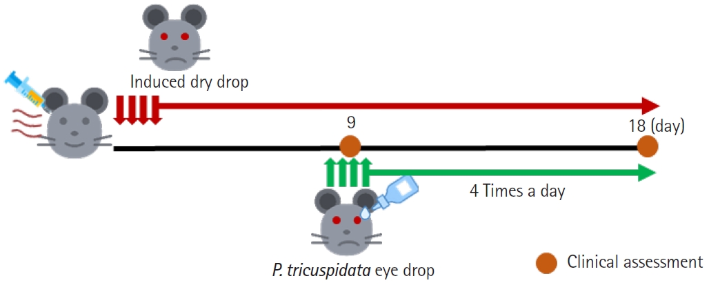
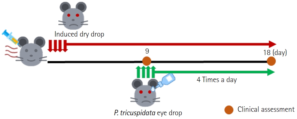
The animal experiments were divided into four groups, and the experimental procedure was conducted over a period of 18 days (Table 1). After 9 days, PT (100, 500 μg/mL) was administered concurrently with the induction of DED. PT was administered as eye drops four times daily, with 5 μL applied to each eye. Three assessments were performed: tear volume, corneal fluorescein staining score, and tear break-up time (TBUT) measurements. The assessments were conducted on day 9 of the experiment to confirm the induction of DED and again on day 18 to evaluate the effects of PT. After the experiment, eyes were removed, and tissue staining was performed (Fig. 1).
Tear volume measurements
Tear volume was assessed using phenol red-impregnated cotton threads (Zone-Quick, Showa Yakuhin Kako Co.) following previously described methods [17]. In brief, the cotton threads were placed at the lateral canthus for 20 seconds to measure tear volume, quantified by the length of the thread (mm) that turned red. A standard curve was generated to convert the distance traveled by the red dye into tear volume.
Corneal fluorescein staining score
Corneal fluorescein staining of eyes was conducted using 0.5% proxymetacaine hydrochloride (Alcaine, Alcon Lab Inc.). Next, the eyes were evaluated for corneal staining under a slit-lamp biomicroscope according to the Oxford schema [18]. The degree of corneal staining was graded as follows: 0 for normal; 1 to 2 for mild to moderate; and ≥3 for severe [19].
Evaluation of TBUT
TBUT was used to assess DED. First, fluorescein was instilled into the tear film of mice and observed under broad cobalt blue illumination. TBUT was quantified as the duration (in seconds) between the final blink and the appearance of the first dry spot in the tear film, as observed through successive slit-lamp photos over time. Each measurement was repeated three times, and the average value reported.
Histological and immunohistochemical analyses
The eyes were removed from mice before immunostaining. On the following day, the eyes were transferred to 30% sucrose (Sigma-Aldrich) overnight at 4 °C. Tissue sections (10-µm-thick) were cut using a cryostat and thaw-mounted onto histological slides. The frozen sections were stained using an Alcian Blue Staining Kit (Abcam) according to the manufacturer’s instructions. Briefly, the tissue was washed with distilled water and incubated with an acetic acid solution for 3 minutes. Subsequently, the tissue was stained with Alcian blue solution at room temperature for 30 minutes, followed by washing and staining with safranin O solution (Dako) for 5 minutes. Alcian blue stains acidic mucin present in goblet cells.
Immunohistochemical analysis was conducted following protocols used in previous studies. The sections were washed twice with 1× phosphate-buffered saline for 5 minutes each and subsequently blocked with 5% BSA for 1 hour. Following blocking, sections were incubated overnight at 4 °C with primary antibodies (IL-6, IL-1β, and tumor necrosis factor-α [TNF-α]). The next day, sections were incubated with biotinylated secondary antibody (goat anti-rabbit) (Vector Laboratories) diluted at 1:500 in BSA for 1 hour at room temperature. Sections were further treated with avidin-biotin complex kit (Vector Laboratories) for 1 hour, followed by incubation with 3,3′-diaminobenzidine peroxidase substrate kit (DAB) (Vector Laboratories). Counterstaining was performed using Meyer’s hematoxylin (Dako). Stained sections were examined using a DXM1200F microscope (Nikon), and the processed images were analyzed for cell counts using the ImageJ software ver. 1.50i (National Institutes of Health).
Statistical analysis
Data were analyzed using GraphPad Prism software ver. 7.0 (GraphPad) and are presented as the mean±standard error of the mean. Statistical significance was determined using one-way analysis of variance followed by Tukey post hoc test. A P-value <0.05 was considered statistically significant.
Measurement of DPPH radical scavenging activity
Free radicals readily oxidize lipids and proteins. DPPH is a relatively stable free radical [20]. The DPPH assay is widely used to assess short-term antioxidant activity. The DPPH radical scavenging activity of PT was compared with that of the synthetic antioxidant Trolox, indicating a concentration-dependent increase in all concentrations of PT (Table 2). Consequently, PT exhibited superior antioxidant effects compared to Trolox.
Effect of PT on H2O2-exposed WKD cells
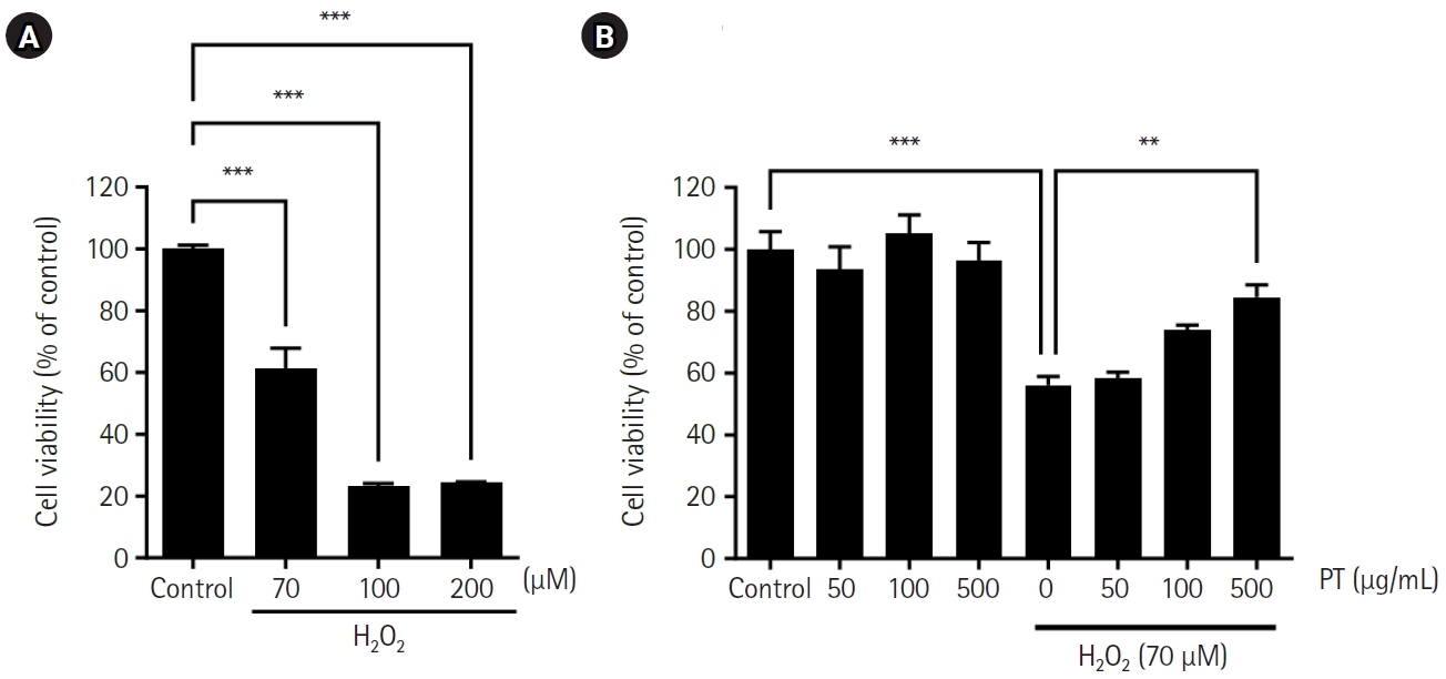
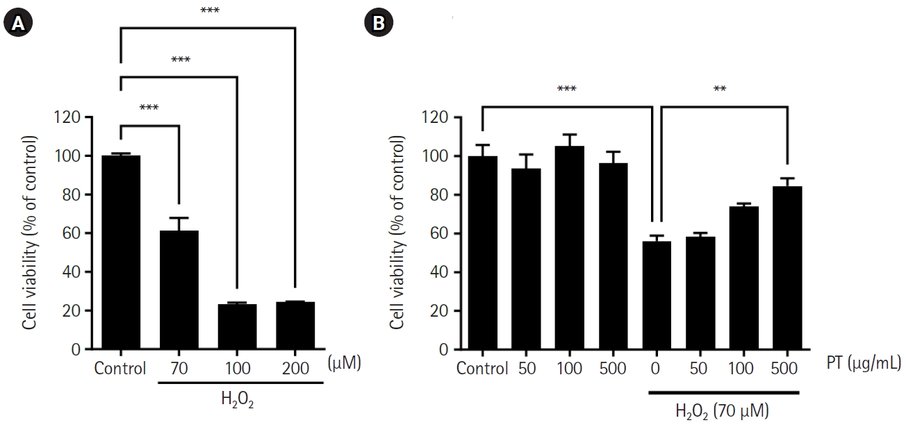
To assess the cytotoxic effects of H2O2 on WKD cells, MTT assays were conducted. The viability of WKD cells exposed to H2O2 decreased and the IC50 value was 70 μM (Fig. 2A). Consequently, for subsequent experiments, a concentration of 70 μM was used to evaluate H2O2-induced apoptosis. Fig. 2B presents the effects of PT on WKD cell apoptosis induced by 70 μM H2O2. Following treatment with 50, 100, and 500 μg/mL PT indicating no toxicity to cells. Notably, PT demonstrated therapeutic effects on H2O2-exposed WKD cells.
Effects of PT on ROS levels in H2O2-exposed WKD cells
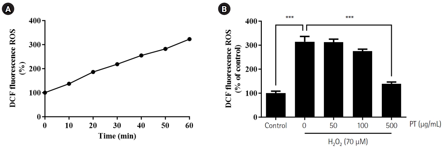
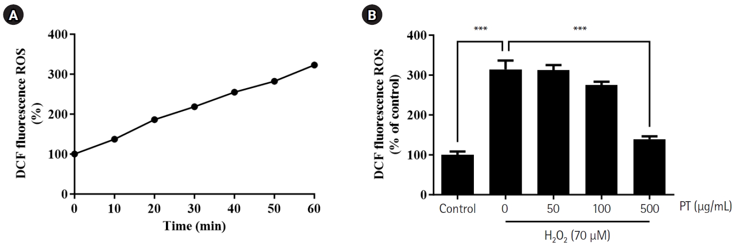
Under conditions of oxidative stress, there was a significant increase in ROS production. After exposure to 70 μM H2O2 alone, cellular ROS levels increased over time (Fig. 3A). However, pretreatment of cells with PT led to a reduction in ROS production. Cells pretreated with PT at 500 μg/mL and then exposed to H2O2 exhibited ROS levels comparable to those of control cells (Fig. 3B). Thus, PT can inhibit the H2O2-induced increase in ROS production in WKD cells.
Effects of PT on mitogen-activated protein kinase phosphorylation in H2O2-exposed WKD cells
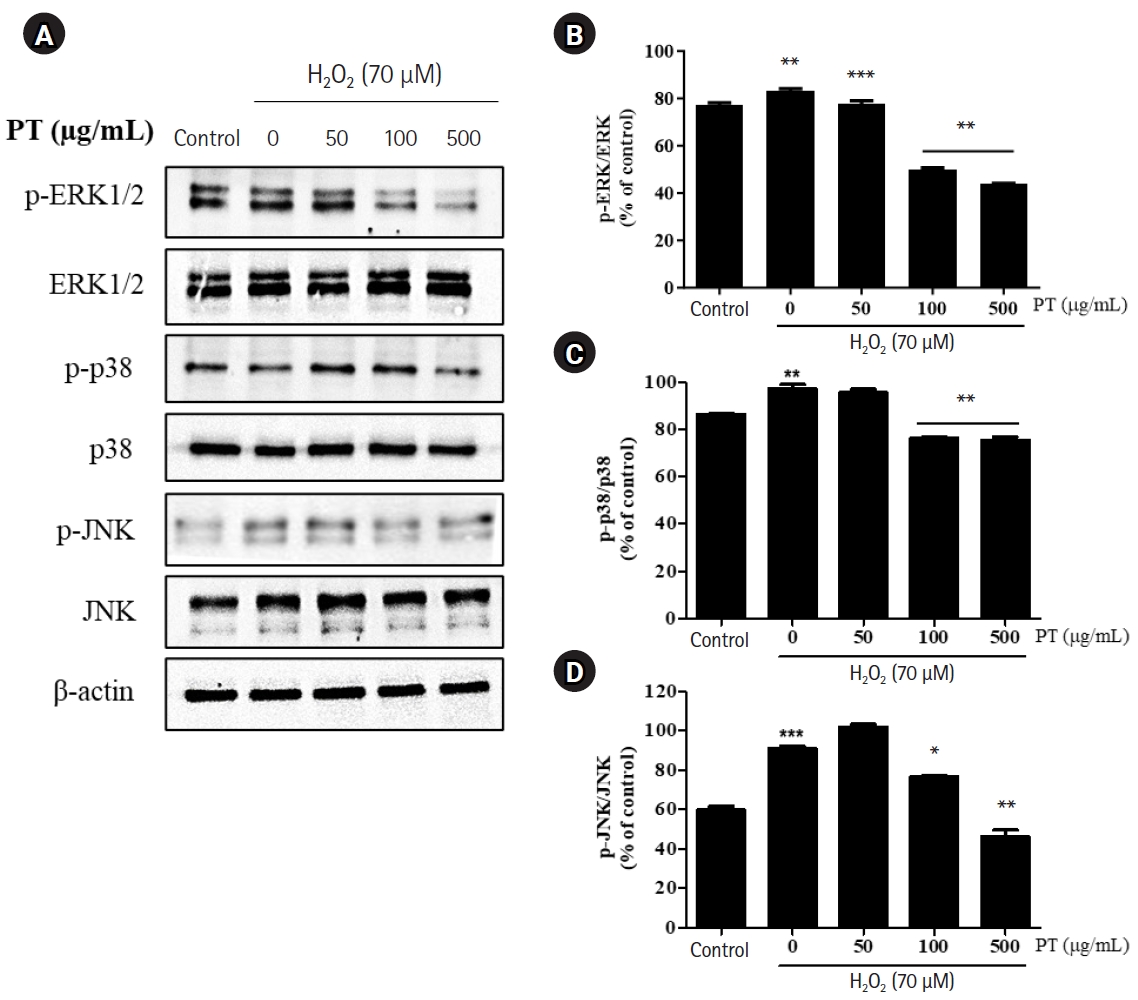
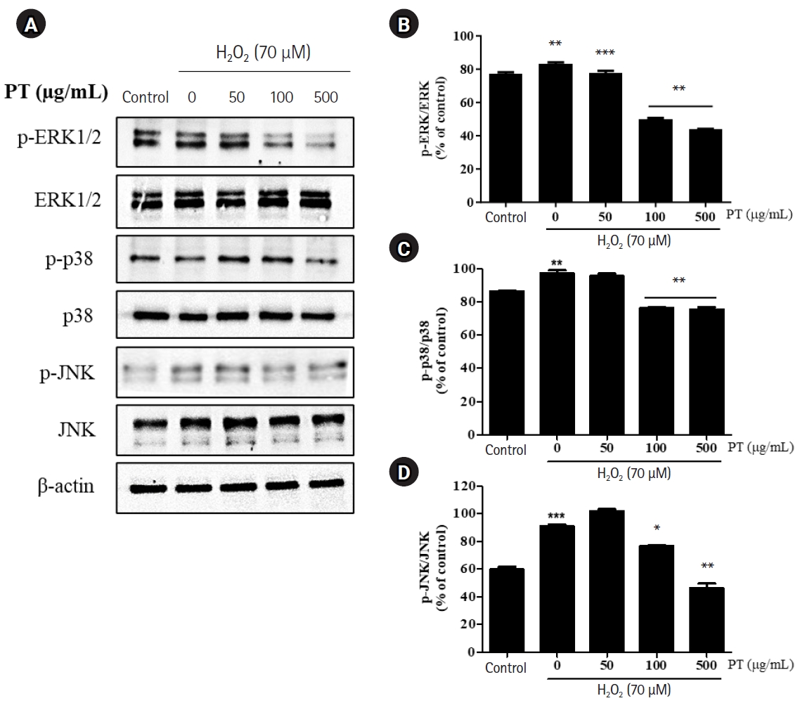
We examined the activation of p38, ERK1/2, and JNK in the mitogen-activated protein kinase (MAPK) family to elucidate the therapeutic mechanism of PT in WKD cells. The effects of PT on the MAPK pathway were analyzed via Western blotting. The original Western blot images were shown in Fig. S1. Exposure to 70 μM H2O2 increased (compared to the control group) the levels of p-p38/p38, p-JNK/JNK, and p-ERK1/2/ERK1/2 However, in the 50, 100, and 500 μg/mL PT-treated groups, the levels of p-p38/p38, p-JNK/JNK, and p-ERK1/2/ERK1/2 decreased (compared to the H2O2-exposed group). These findings suggest that the effects of PT treatment in reducing antioxidant consumption and enhancing antioxidant levels may occur via the MAPK signaling pathway to protect WKD cells from oxidative stress (Fig. 4).
Effects of PT on SOD, CAT, and MDA levels in H2O2-exposed WKD cells
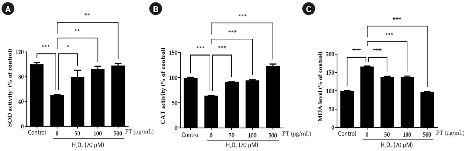
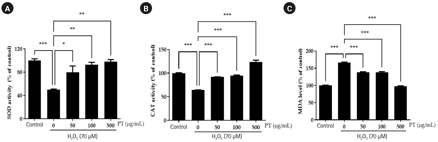
The effects of PT pretreatment on 70 μM H2O2-exposed WKD cells were evaluated based on CAT (Fig. 5A) and SOD (Fig. 5B) activities, and MDA levels (Fig. 5C). In H2O2-exposed cells, the activities of SOD and CAT were reduced (compared to the control); MDA levels increased. Treatment with 50, 100, and 500 μg/mL PT increased (compared to the H2O2-exposed group) SOD, CAT activity and reduced MDA levels. Notably, 500 μg/mL PT increased SOD and CAT activity to levels comparable to those of the control group. These results suggest that PT exhibits strong antioxidant activity.
Effects of PT on pro-inflammatory cytokine production in H2O2-exposed WKD cells
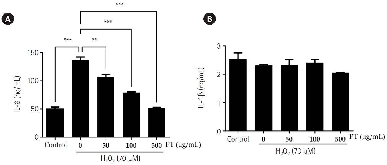
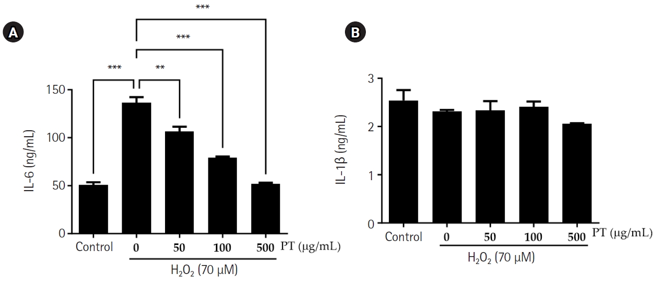
We evaluated the levels of IL-6 and IL-1β in the medium of H2O2-exposed cells to determine the levels of inflammatory cytokines. In H2O2-exposed cells, the IL-6 level increased (compared to the control). Treatment with 50, 100, and 500 μg/mL PT reduced IL-6 production. There was no significant difference in IL-1β levels (Fig. 6).
Effects of PT treatment on an in vivo mouse model of dry eyes
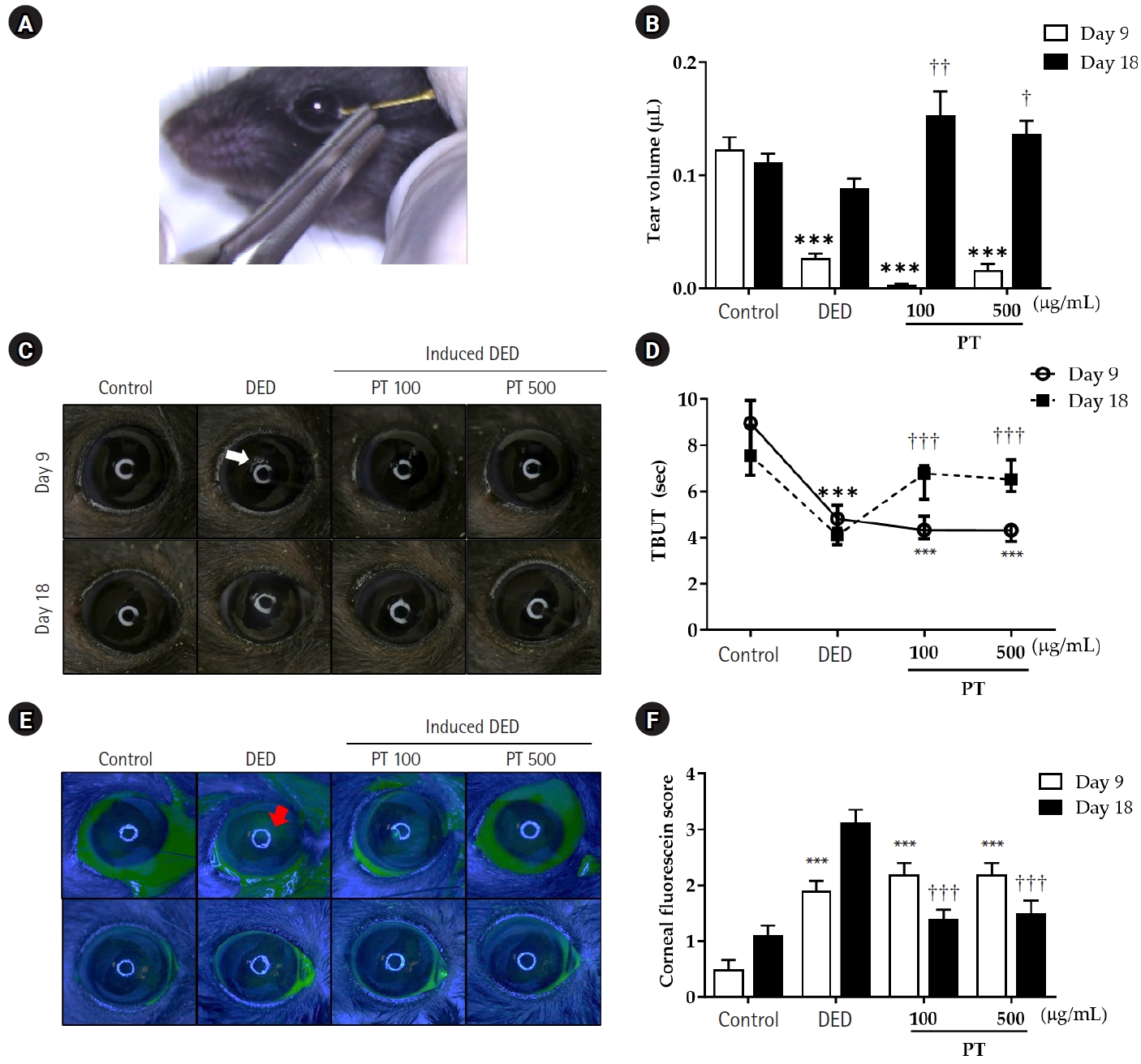
Assessments were conducted at two points: on day 9 after dry eye induction and on day 18 (9 days after PT treatment began). Tear volume, as a typical symptom of dry eye, tear volume reduced significantly in the DED group compared to the control group. After 9 days of PT treatment, tear volume was increased in all groups (Fig. 7B). The PT-treated groups showed recovery to levels similar to the control group. Corneal irregularities, indicative of desiccation stress, appeared as distorted white rings of all groups with induced dry eye (thus, excluding the control group). After 9 days of PT treatment, the PT-treated groups exhibited circular white rings similar to those of the control group (Fig. 7C). On experimental day 9, TBUT in the DED group was significantly decreased, compared to the control group. However, after 9 days of PT treatment, TBUT was higher in the 100 and 500 μg/mL PT groups compared to the DED group (Fig. 7D). Finally, fluorescein staining of the cornea, which increases during dry eye, was significantly increased in the DED group compared to the control group (Fig. 7E). After 9 days of PT treatment, the fluorescein staining score of the DED group was higher than that on day 9; however, the 100 and 500 μg/mL PT groups exhibited significantly less staining than the DED group (Fig. 7F).
Effect of PT on conjunctival goblet cell population in vivo
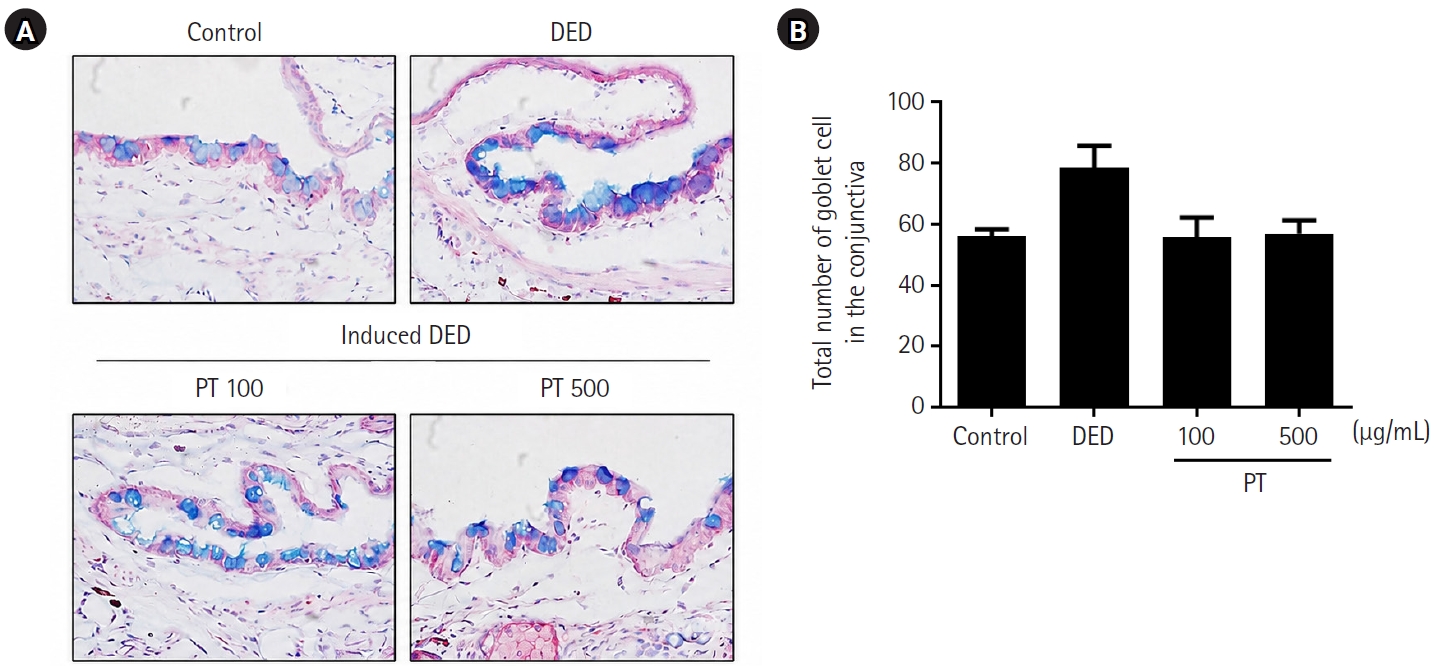
The conjunctiva was stained with Alcian blue, resulting in violet-colored goblet cells. The total number of goblet cells tended to be higher in the DED group than in the control group; however, the difference was not statistically significant. In addition, PT treatment tended to reduce the total number of goblet cells compared with the DED group, although the differences were not statistically significant (Fig. 8).
Effects of PT on inflammatory cytokine expression in the cornea and conjunctiva
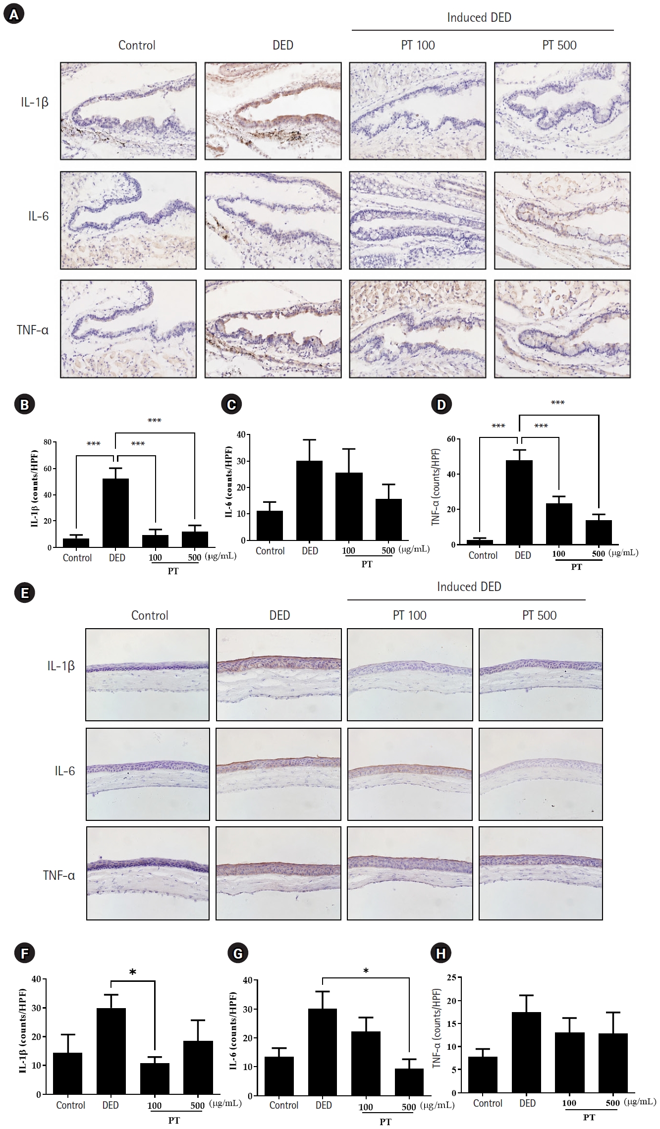
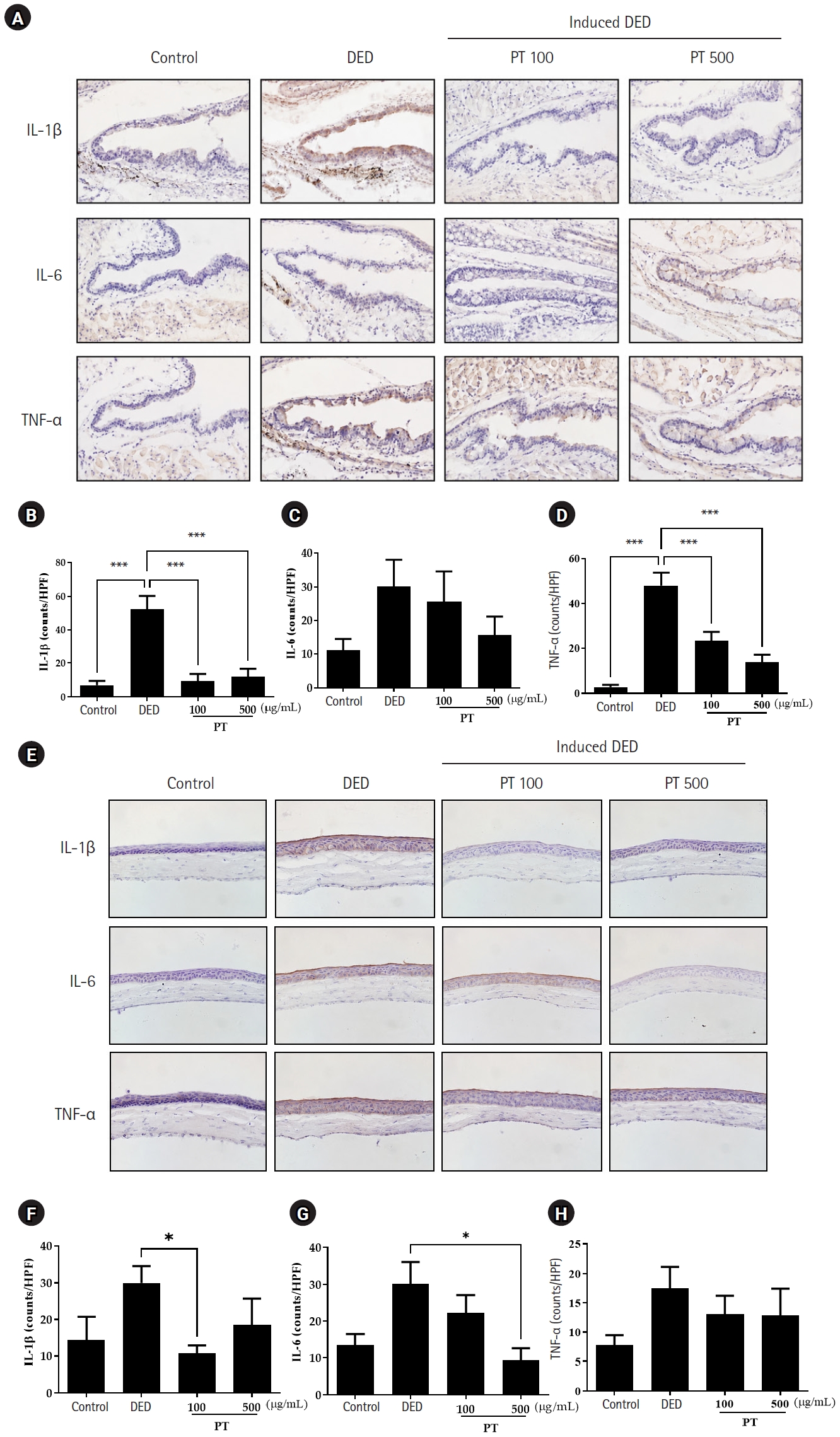
Fig. 9 presents corneal and conjunctiva sections immunostained with antibodies against inflammatory cytokines. The expression levels of IL-1β, IL-6, and TNF-α in the conjunctiva and cornea were higher in the DED group than in the control group. Overall, the expression IL-1, IL-6 and TNF-α was decreased in the PT groups compared to the DED group (Fig. 9).
DED represents a complex condition characterized by disturbances in tear film stability and ocular surface homeostasis, leading to ocular inflammation and potential damage [21]. Oxidative stress, marked by an imbalance between ROS production and elimination, is implicated in its pathogenesis. Therefore, we used WKD cells, a human conjunctival epithelial cell line, to investigate the effects of PT on H2O2-induced oxidative stress.
Current treatments for dry eye often involve anti-inflammatory agents, but these can have side effects with prolonged use [22,23]. Therefore, the development of treatments without side effects is required. Natural extracts have been investigated for DED due to their fewer side effects. Studies have shown that P. tricuspidata has anti-inflammatory and antioxidant properties [24]. In this study, we investigated the effects of PT, a natural antioxidant, on DED models [25]. P. tricuspidata exhibits high antioxidant activity and inhibits aging because of its potent ability to reduce or eliminate free radicals when the DPPH radical scavenging ability is high [26].
In the present study, pretreatment with PT inhibited H2O2-induced apoptosis in WKD cells. Thus, PT inhibits oxidative stress-induced ROS levels. Moreover, members of the MAPK family, such as ERK1/2, JNK, and p38, are crucial regulators of inflammatory cytokine expression and signaling pathways. The MAPK signaling pathway activates cells in response to inflammatory markers, increasing the expression of inflammatory cytokines [27]. These kinases are activated by inflammatory cytokines, such as IL-6 and IL-1β, as well as various stress stimuli to induce biological responses such as inflammation, apoptosis, and cell differentiation [27]. In the present study, H2O2-exposed WKD cells exhibited increased MAPK phosphorylation (compared to the control), which was significantly rescued by PT treatment. Thus, PT regulates MAPK molecules. Cellular oxidative stress can trigger apoptosis due reductions in SOD [28] and CAT [29] activity and increased MDA level [30]. In the present study, PT treatment increased SOD and CAT activities and reduced the MDA level compared to the H2O2-exposed group. Thus, PT can enhance antioxidant enzyme production, resulting in decreased ROS levels, which can potentially prevent oxidative damage of WKD cells. IL-6 and IL-1β are crucial indicators of inflammation, and their expression in conjunctival epithelial cells is increased in DED [15]. Compared to the H2O2-exposed group, PT-treated groups exhibited reduced IL-6 production. Based on the in vitro effects of PT on oxidative stress, we evaluated its efficacy in a mouse model of DED induced by scopolamine administration and a desiccating environment. The tear volume and TBUT were reduced, and fluorescence staining increased, in the DED mouse model; after administration of concentrated PT eye drops, the tear volume and TBUT were increased and fluorescence staining score decreased in mice with DED. In DED, the expression of cytokines causing tissue inflammation is increased [31]. After removal of the mouse eyes, evaluation of IL-6, IL-1β, and TNF-α levels in the cornea and tissue staining of the conjunctiva revealed a significant reduction in inflammatory cytokine levels in the PT-treated group compared to the DED group.
Conclusion
In summary, this study showed that PT protects the ocular surface in the DED model by reducing ROS production and oxidative damage. Therefore, it is expected to be developed as an antioxidant with fewer side effects as a natural substance in the future through more systematic studies using PT. In other words, PT seems to be a potential therapeutic agent for ocular surface diseases such as DED.

Author contributions

Conceptualization: KJC. Data curation: HG. Formal analysis: HG. Visualization: HG, CHO. Writing – original draft: HG. Writing – review & editing: CHO, KJC. Final approval of the manuscript: all authors.

Conflicts of interest

No potential conflict of interest relevant to this article was reported.

Funding

None.

Data availability

The data that support the findings of the current study may be requested from the corresponding author upon reasonable request.

Supplementary materials can be found via https://doi.org/10.63375/icrs.26.005.
Fig. S1
Image of original uncropped blots used for preparation of Fig. 3A for p-ERK1/2, ERK1/2, p-p38, p38, p-JNK, JNK, and β-actin Western blots. PT, Parthenocissus tricuspidata growing on pine; p-ERK1/2, phosphorylated extracellular signal-regulated kinase 1/2; p-p38, phosphorylated p38; p-JNK, phosphorylated c-Jun N-terminal kinase.
icrs-26-005-Supplementary-Fig-1.pdf
Fig. 1.
Experimental schedule for the dry eye mouse model and Parthenocissus tricuspidata treatment. To induce dry eye disease, scopolamine was administered subcutaneously four times daily, followed by desiccation stress for 18 hr/day. Beginning on day 9, P. tricuspidata extract was administered as eye drops four times daily. Assessments were performed on days 9 and 18 after the start of the experiment.
icrs-26-005f1.jpg
Fig. 2.
Effect of Parthenocissus tricuspidata growing on pine (PT) extract on cell viability in H2O2-induced Wong-Kilbourne derivative of Chang conjunctival cells. (A) Cell viability was determined using the MTT assay after treatment with various H2O2 concentrations. (B) Cells were pretreated with PT for 1 hour before the addition of 70 μM H2O2 and were then incubated for 24 hours. Results are expressed as a percentage of control values and are reported as the mean±standard error of the mean. MTT, 3-(4,5-dimethylthiazol-2-yl)-2,5-diphenyltetrazolium bromide. Statistically significant (**P<0.01, ***P<0.001).
icrs-26-005f2.jpg
Fig. 3.
Effects of Parthenocissus tricuspidata growing on pine (PT) extract on reactive oxygen species (ROS) production in Wong-Kilbourne derivative of Chang conjunctival cells. (A) H2O2 increased ROS activity in a time-dependent manner. (B) PT suppressed H2O2-induced ROS generation in a dose-dependent manner. Values are expressed as a percentage of control values and are reported as the mean±standard error of the mean. DCF, 2′,7′-dichlorofluorescein. ***Statistically significant (P<0.001).
icrs-26-005f3.jpg
Fig. 4.
Effects of Parthenocissus tricuspidata growing on pine (PT) extract on mitogen-activated protein kinase (MAPK) phosphorylation in H2O2-induced Wong-Kilbourne derivative of Chang conjunctival cells. (A) Effects of PT on MAPK expression. Quantitative analysis of p-ERK1/2/ERK1/2 (B), p-p38/p38 (C), and p-JNK/JNK (D). MAPK phosphorylation was evaluated by Western blot analysis. Values are expressed as a percentage of control values and are reported as the mean±standard error of the mean. PT, Parthenocissus tricuspidata growing on pine; p-ERK1/2, phosphorylated extracellular signal-regulated kinase 1/2; p-p38, phosphorylated p38; p-JNK, phosphorylated c-Jun N-terminal kinase. Statistically significant (*P<0.05, **P<0.01, ***P<0.001).
icrs-26-005f4.jpg
Fig. 5.
Effects of Parthenocissus tricuspidata growing on pine (PT) extract on antioxidant enzyme activities and an oxidation marker in Wong-Kilbourne derivative of Chang conjunctival cells. Activities of superoxide dismutase (SOD) (A), catalase (CAT) (B), as well as malondialdehyde (MDA) levels (C), were measured by enzyme-linked immunosorbent assay. Values are expressed as a percentage of control values and are reported as the mean±standard error of the mean. Statistically significant (*P<0.05, **P<0.01, ***P<0.001).
icrs-26-005f5.jpg
Fig. 6.
Effects of Parthenocissus tricuspidata growing on pine (PT) extract on pro-inflammatory cytokine production in Wong-Kilbourne derivative of Chang conjunctival cells. Levels of interleukin (IL)-6 (A) and IL-1β (B) in cell culture supernatants were determined by enzyme-linked immunosorbent assay. Values are reported as the mean±standard error of the mean. Statistically significant (**P<0.01, ***P<0.001).
icrs-26-005f6.jpg
Fig. 7.
Clinical effects of Parthenocissus tricuspidata growing on pine (PT) extract treatment on scopolamine-induced dry eye in mice. (A) Representative image of tear volume measurement in mice using Zone-Quick. (B) Tear volume in mice. On day 9, groups exposed to desiccation stress showed decreased tear volume compared with the control group. On day 18, tear volume was significantly increased in the PT-treated groups compared with the dry eye disease (DED) group. (C) Eye images showing the effects of PT treatment on corneal surface irregularities. Images were obtained using a microscope. The white arrow indicates a distorted white ring. (D) Tear break-up time (TBUT) in mice. TBUT was significantly reduced in the DED group compared with the control group, whereas PT treatment significantly increased TBUT compared with the DED group. Representative photographs (E) and scores (F) of corneal fluorescein staining in the control, DED, and PT-treated groups on days 9 and 18. On day 18, corneal fluorescein staining scores were lower in the PT-treated groups than in the DED group. The red arrow indicates a fluorescein-stained region in a DED eye. Values are reported as the mean±standard error of the mean. ***P<0.001 compared with the control group on experimental day 9; P<0.05, ††P<0.01, and †††P<0.001 compared with the DED group on experimental day 18.
icrs-26-005f7.jpg
Fig. 8.
Effects of Parthenocissus tricuspidata growing on pine (PT) extract treatment on conjunctival goblet cell number. (A) Goblet cells in the conjunctiva are stained violet with Alcian blue after PT treatment following 8 days of desiccation stress (×100). (B) Number of goblet cells in the control, dry eye disease (DED), and PT-treated groups. Quantitative data are reported as the mean±standard error of the mean.
icrs-26-005f8.jpg
Fig. 9.
Effects of Parthenocissus tricuspidata growing on pine (PT) extract on inflammatory cytokine expression in the conjunctiva and cornea. (A, E) Conjunctival and corneal sections from the control, dry eye disease (DED), 100 μg/mL PT, and 500 μg/mL PT groups immunostained for interleukin (IL)-1β, IL-6, and tumor necrosis factor (TNF)-α. Images were captured using a microscope and camera (×100). IL-1β (B, F), IL-6 (C, G), and TNF-α (D, H) expression in the experimental groups, expressed as percentages of the control group and quantified using ImageJ software. Quantitative data are reported as the mean±standard error of the mean. HPF, high-power fields. Statistically significant (*P<0.05, ***P<0.001).
icrs-26-005f9.jpg
Table 1.
The mice were divided into four groups according to the treatment as follows
Group Treatment
Control Control (not induced dry eye)
DED Induced dry eye
PT 100 Induced dry eye and PT 100 μg/mL
PT 500 Induced dry eye and PT 500 μg/mL

DED, dry eye disease; PT, Parthenocissus tricuspidata growing on pine.

Table 2.
DPPH radical scavenging activity of PT extracts
Compound DPPH radical scavenging (%)
Trolox 1.94±0.40
PT 50 μg/mL 60.40±5.18***
PT 100 μg/mL 63.93±1.93***
PT 500 μg/mL 64.92±2.06***

Values are presented as mean±standard error of the mean.

DPPH, 2,2-diphenyl-1-picrylhydrazyl; PT, Parthenocissus tricuspidata growing on pine.

***P<0.001 compared to with the Trolox.

  • 1. Stern ME, Pflugfelder SC. Inflammation in dry eye. Ocul Surf 2004;2:124‒30. ArticlePubMed
  • 2. Wenk J, Brenneisen P, Meewes C, et al. UV-induced oxidative stress and photoaging. Curr Probl Dermatol 2001;29:83‒94. ArticlePubMed
  • 3. Lee SJ, Kim EK, Hwang JW, et al. Purification and characterisation of an antioxidative peptide from enzymatic hydrolysates of duck processing by-products. Food Chem 2010;123:216‒20. Article
  • 4. Imamura Y, Noda S, Hashizume K, et al. Drusen, choroidal neovascularization, and retinal pigment epithelium dysfunction in SOD1-deficient mice: a model of age-related macular degeneration. Proc Natl Acad Sci U S A 2006;103:11282‒7. ArticlePubMedPMC
  • 5. Noecker R. Effects of common ophthalmic preservatives on ocular health. Adv Ther 2001;18:205‒15. ArticlePubMedPDF
  • 6. Goto E, Yagi Y, Matsumoto Y, Tsubota K. Impaired functional visual acuity of dry eye patients. Am J Ophthalmol 2002;133:181‒6. ArticlePubMed
  • 7. Musch DC, Sugar A, Meyer RF. Demographic and predisposing factors in corneal ulceration. Arch Ophthalmol 1983;101:1545‒8. ArticlePubMed
  • 8. Perez-Garcia F, Marin E, Canigueral S, Adzet T. Anti-inflammatory action of Pluchea sagittalis: involvement of an antioxidant mechanism. Life Sci 1996;59:2033‒40. ArticlePubMed
  • 9. Tuzun A, Erdil A, Inal V, et al. Oxidative stress and antioxidant capacity in patients with inflammatory bowel disease. Clin Biochem 2002;35:569‒72. ArticlePubMed
  • 10. Lee SJ, Jung DS, Bu HJ, et al. Screening of the tyrosinase inhibition and hyaluronidase inhibition activities, and radical scavenging effects using plants in Cheju. Korean J Pharmacogn 2001;32:175‒80.
  • 11. Marquina MA, Corao GM, Araujo L, et al. Hyaluronidase inhibitory activity from the polyphenols in the fruit of blackberry (Rubus fruticosus B.). Fitoterapia 2002;73:727‒9. ArticlePubMed
  • 12. Lee T. Coloured flora of Korea. Vol Ⅰ․ Hyang-Mun Publishing Co.; 2003.
  • 13. Cho M. Coloured woody plants of Korea. Academic Inc.; 1989. p. 498.
  • 14. Albani KA. Practices, attitudes and perceived benefits of supplement use in patients in phase II cardiac rehabilitation [master's thesis]. Texas Woman’s University; 1999.
  • 15. Alessio HM, Hagerman AE, Fulkerson BK, et al. Generation of reactive oxygen species after exhaustive aerobic and isometric exercise. Med Sci Sports Exerc 2000;32:1576‒81. ArticlePubMed
  • 16. Halliwell B, Gutteridge JM. Free radicals in biology and medicine. Oxford University Press; 2015.
  • 17. Balboni GC, Brandi ML, Zonefrati R, Repice F. Effects of He-Ne/IR laser irradiation on two lines of normal human fibroblasts in vitro. Arch Ital Anat Embriol 1986;91:179‒88. PubMed
  • 18. Bron AJ, Evans VE, Smith JA. Grading of corneal and conjunctival staining in the context of other dry eye tests. Cornea 2003;22:640‒50. ArticlePubMed
  • 19. Labbe A, Terry O, Brasnu E, et al. Tear film osmolarity in patients treated for glaucoma or ocular hypertension. Cornea 2012;31:994‒9. ArticlePubMed
  • 20. Tsubota K, Goto E, Fujita H, et al. Treatment of dry eye by autologous serum application in Sjögren’s syndrome. Br J Ophthalmol 1999;83:390‒5. ArticlePubMedPMC
  • 21. Lemp MA, Foulks GN. The definition and classification of dry eye disease: report of the Definition and Classification Subcommittee of the International Dry Eye Workshop (2007). Ocul Surf 2007;5:75‒92. ArticlePubMed
  • 22. Colligris B, Alkozi HA, Pintor J. Recent developments on dry eye disease treatment compounds. Saudi J Ophthalmol 2014;28:19‒30. ArticlePubMedPMC
  • 23. Finkel T. Oxygen radicals and signaling. Curr Opin Cell Biol 1998;10:248‒53. ArticlePubMed
  • 24. Jo NR, Park MA, Chae KY, et al. Cellular protective and antioxidative acivities of Parthenocissus tricuspidata stem extracts. J Soc Cosmet Sci Korea 2012;38:225‒36. Article
  • 25. Jeong YS, Jung HK, Youn KS, Kim MO, Hong JH. Physiological activities of the hot water extract from Eriobotrya japonica Lindl. J Korean Soc Food Sci Nutr 2009;38:977‒82. Article
  • 26. Aoshima H, Tsunoue H, Koda H, Kiso Y. Aging of whiskey increases 1,1-diphenyl-2-picrylhydrazyl radical scavenging activity. J Agric Food Chem 2004;52:5240‒4. ArticlePubMed
  • 27. Yang H, Oh KH, Yoo YC. Anti-inflammatory effect of hot water extract of aronia fruits in LPS-stimulated RAW 264.7 macrophages. J Korean Soc Food Sci Nutr 2015;44:7‒13. Article
  • 28. Liu X, Ma Y, Wei X, Fan T. Neuroprotective effect of licochalcone A against oxygen‐glucose deprivation/reperfusion in rat primary cortical neurons by attenuating oxidative stress injury and inflammatory response via the SIRT1/Nrf2 pathway. J Cell Biochem 2018;119:3210‒9. ArticlePubMedPDF
  • 29. Jiang WD, Tang RJ, Liu Y, et al. Impairment of gill structural integrity by manganese deficiency or excess related to induction of oxidative damage, apoptosis and dysfunction of the physical barrier as regulated by NF-κB, caspase and Nrf2 signaling in fish. Fish Shellfish Immunol 2017;70:280‒92. ArticlePubMed
  • 30. Tan X, Liu B, Lu J, et al. Dietary luteolin protects against HgCl2-induced renal injury via activation of Nrf2-mediated signaling in rat. J Inorg Biochem 2018;179:24‒31. ArticlePubMed
  • 31. Stevenson W, Chauhan SK, Dana R. Dry eye disease: an immune-mediated ocular surface disorder. Arch Ophthalmol 2012;130:90‒100. ArticlePubMedPMC

Figure & Data

References

    Citations

    Citations to this article as recorded by  

      • ePub LinkePub Link
      • Cite
        CITE
        export Copy Download
        Close
        Download Citation
        Download a citation file in RIS format that can be imported by all major citation management software, including EndNote, ProCite, RefWorks, and Reference Manager.

        Format:
        • RIS — For EndNote, ProCite, RefWorks, and most other reference management software
        • BibTeX — For JabRef, BibDesk, and other BibTeX-specific software
        Include:
        • Citation for the content below
        Effects of extract of Parthenocissus tricuspidata living on pine in a nonclinical model of dry eye disease
        Insights Cataract Refract Surg. 2026;11(2):56-68.   Published online June 30, 2026
        Close
      • XML DownloadXML Download
      Figure
      • 0
      • 1
      • 2
      • 3
      • 4
      • 5
      • 6
      • 7
      • 8
      We recommend
      Effects of extract of Parthenocissus tricuspidata living on pine in a nonclinical model of dry eye disease
      Image Image Image Image Image Image Image Image Image
      Fig. 1. Experimental schedule for the dry eye mouse model and Parthenocissus tricuspidata treatment. To induce dry eye disease, scopolamine was administered subcutaneously four times daily, followed by desiccation stress for 18 hr/day. Beginning on day 9, P. tricuspidata extract was administered as eye drops four times daily. Assessments were performed on days 9 and 18 after the start of the experiment.
      Fig. 2. Effect of Parthenocissus tricuspidata growing on pine (PT) extract on cell viability in H2O2-induced Wong-Kilbourne derivative of Chang conjunctival cells. (A) Cell viability was determined using the MTT assay after treatment with various H2O2 concentrations. (B) Cells were pretreated with PT for 1 hour before the addition of 70 μM H2O2 and were then incubated for 24 hours. Results are expressed as a percentage of control values and are reported as the mean±standard error of the mean. MTT, 3-(4,5-dimethylthiazol-2-yl)-2,5-diphenyltetrazolium bromide. Statistically significant (**P<0.01, ***P<0.001).
      Fig. 3. Effects of Parthenocissus tricuspidata growing on pine (PT) extract on reactive oxygen species (ROS) production in Wong-Kilbourne derivative of Chang conjunctival cells. (A) H2O2 increased ROS activity in a time-dependent manner. (B) PT suppressed H2O2-induced ROS generation in a dose-dependent manner. Values are expressed as a percentage of control values and are reported as the mean±standard error of the mean. DCF, 2′,7′-dichlorofluorescein. ***Statistically significant (P<0.001).
      Fig. 4. Effects of Parthenocissus tricuspidata growing on pine (PT) extract on mitogen-activated protein kinase (MAPK) phosphorylation in H2O2-induced Wong-Kilbourne derivative of Chang conjunctival cells. (A) Effects of PT on MAPK expression. Quantitative analysis of p-ERK1/2/ERK1/2 (B), p-p38/p38 (C), and p-JNK/JNK (D). MAPK phosphorylation was evaluated by Western blot analysis. Values are expressed as a percentage of control values and are reported as the mean±standard error of the mean. PT, Parthenocissus tricuspidata growing on pine; p-ERK1/2, phosphorylated extracellular signal-regulated kinase 1/2; p-p38, phosphorylated p38; p-JNK, phosphorylated c-Jun N-terminal kinase. Statistically significant (*P<0.05, **P<0.01, ***P<0.001).
      Fig. 5. Effects of Parthenocissus tricuspidata growing on pine (PT) extract on antioxidant enzyme activities and an oxidation marker in Wong-Kilbourne derivative of Chang conjunctival cells. Activities of superoxide dismutase (SOD) (A), catalase (CAT) (B), as well as malondialdehyde (MDA) levels (C), were measured by enzyme-linked immunosorbent assay. Values are expressed as a percentage of control values and are reported as the mean±standard error of the mean. Statistically significant (*P<0.05, **P<0.01, ***P<0.001).
      Fig. 6. Effects of Parthenocissus tricuspidata growing on pine (PT) extract on pro-inflammatory cytokine production in Wong-Kilbourne derivative of Chang conjunctival cells. Levels of interleukin (IL)-6 (A) and IL-1β (B) in cell culture supernatants were determined by enzyme-linked immunosorbent assay. Values are reported as the mean±standard error of the mean. Statistically significant (**P<0.01, ***P<0.001).
      Fig. 7. Clinical effects of Parthenocissus tricuspidata growing on pine (PT) extract treatment on scopolamine-induced dry eye in mice. (A) Representative image of tear volume measurement in mice using Zone-Quick. (B) Tear volume in mice. On day 9, groups exposed to desiccation stress showed decreased tear volume compared with the control group. On day 18, tear volume was significantly increased in the PT-treated groups compared with the dry eye disease (DED) group. (C) Eye images showing the effects of PT treatment on corneal surface irregularities. Images were obtained using a microscope. The white arrow indicates a distorted white ring. (D) Tear break-up time (TBUT) in mice. TBUT was significantly reduced in the DED group compared with the control group, whereas PT treatment significantly increased TBUT compared with the DED group. Representative photographs (E) and scores (F) of corneal fluorescein staining in the control, DED, and PT-treated groups on days 9 and 18. On day 18, corneal fluorescein staining scores were lower in the PT-treated groups than in the DED group. The red arrow indicates a fluorescein-stained region in a DED eye. Values are reported as the mean±standard error of the mean. ***P<0.001 compared with the control group on experimental day 9; †P<0.05, ††P<0.01, and †††P<0.001 compared with the DED group on experimental day 18.
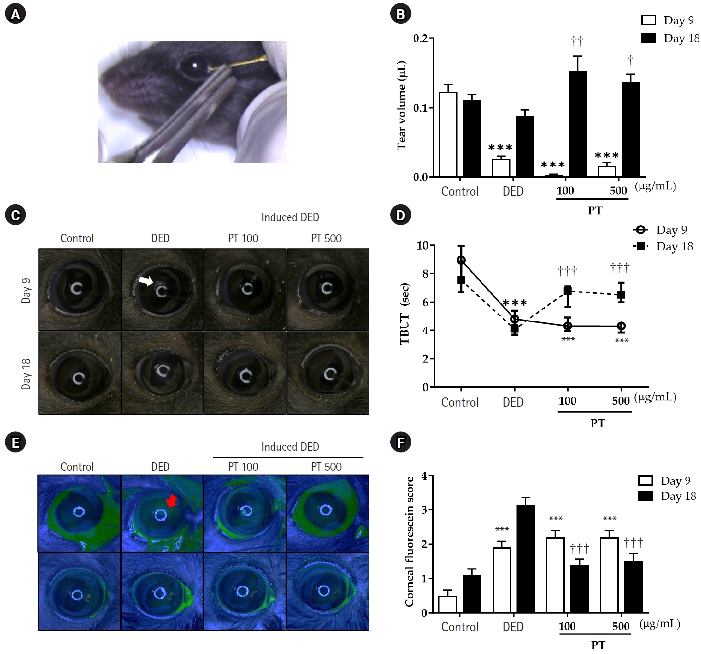
      Fig. 8. Effects of Parthenocissus tricuspidata growing on pine (PT) extract treatment on conjunctival goblet cell number. (A) Goblet cells in the conjunctiva are stained violet with Alcian blue after PT treatment following 8 days of desiccation stress (×100). (B) Number of goblet cells in the control, dry eye disease (DED), and PT-treated groups. Quantitative data are reported as the mean±standard error of the mean.
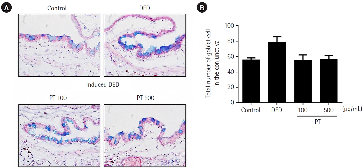
      Fig. 9. Effects of Parthenocissus tricuspidata growing on pine (PT) extract on inflammatory cytokine expression in the conjunctiva and cornea. (A, E) Conjunctival and corneal sections from the control, dry eye disease (DED), 100 μg/mL PT, and 500 μg/mL PT groups immunostained for interleukin (IL)-1β, IL-6, and tumor necrosis factor (TNF)-α. Images were captured using a microscope and camera (×100). IL-1β (B, F), IL-6 (C, G), and TNF-α (D, H) expression in the experimental groups, expressed as percentages of the control group and quantified using ImageJ software. Quantitative data are reported as the mean±standard error of the mean. HPF, high-power fields. Statistically significant (*P<0.05, ***P<0.001).
      Effects of extract of Parthenocissus tricuspidata living on pine in a nonclinical model of dry eye disease
      Group Treatment
      Control Control (not induced dry eye)
      DED Induced dry eye
      PT 100 Induced dry eye and PT 100 μg/mL
      PT 500 Induced dry eye and PT 500 μg/mL
      Compound DPPH radical scavenging (%)
      Trolox 1.94±0.40
      PT 50 μg/mL 60.40±5.18***
      PT 100 μg/mL 63.93±1.93***
      PT 500 μg/mL 64.92±2.06***
      Table 1. The mice were divided into four groups according to the treatment as follows

      DED, dry eye disease; PT, Parthenocissus tricuspidata growing on pine.

      Table 2. DPPH radical scavenging activity of PT extracts

      Values are presented as mean±standard error of the mean.

      DPPH, 2,2-diphenyl-1-picrylhydrazyl; PT, Parthenocissus tricuspidata growing on pine.

      P<0.001 compared to with the Trolox.


      Insights Cataract Refract Surg : Insights in Cataract and Refractive Surgery
      Close layer
      TOP