
 KSCRS

Current issue
- Page Path
- HOME > Browse articles > Current issue
- Volume 11(3); October 2026
-


Review Article
- Premium intraocular lens selection in glaucoma: an approach based on visual function and disease characteristics
- Yeoun Sook Chun
- Insights Cataract Refract Surg 2026;11(3):69-82. Published online October 29, 2026
- DOI: https://doi.org/10.63375/icrs.26.008
-
 Abstract
 PDF
 ePub
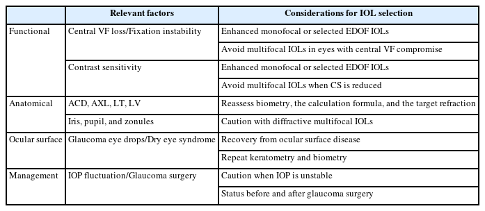
- The increasing global incidence of cataracts and glaucoma, together with rising patient expectations, is leading to a steady increase in premium intraocular lens (IOL) implantation. However, concerns remain about the use of premium IOLs in eyes with glaucoma. Selecting an IOL for a patient with glaucoma requires comprehensive consideration of central visual field defects, contrast sensitivity, fixation stability, anterior segment anatomy, ocular surface disease, intraocular pressure control, topical medication use, previous or anticipated glaucoma surgery, and the lifelong risk of glaucoma progression. For glaucoma suspects and patients with ocular hypertension or early, stable open-angle glaucoma, enhanced monofocal IOLs may help improve intermediate vision while maintaining distance visual acuity and preserving contrast sensitivity. Extended depth-of-focus IOLs may also be considered for selected patients with glaucoma and preserved central visual function. Toric IOLs have generally yielded favorable outcomes, but their use should be considered only when astigmatism is regular and reproducible and capsular–zonular support is stable. Multifocal IOL implantation is generally not recommended for patients with glaucoma and visual field defects; it should be considered only in highly selected glaucoma suspects or patients with very early, stable glaucoma, after careful functional assessment and with long-term monitoring. This review aims to systematically summarize considerations for individualized IOL selection and preoperative counseling in patients with glaucoma, integrating functional vulnerability, ocular anatomy, refractive variability, glaucoma subtypes, and the risk of disease progression with the optical characteristics of each IOL design.
- 26 View
- 0 Download

Original Articles
- Comparison of toric intraocular lenses and limbal relaxing incision in extended-depth-of-focus intraocular lens implantation
- Jiyoung Emily Lee, Eun Chul Kim
- Insights Cataract Refract Surg 2026;11(3):83-90. Published online September 23, 2026
- DOI: https://doi.org/10.63375/icrs.26.003
-
Abstract
PDFePub
- Purpose
This study aimed to compare visual and refractive outcomes after toric intraocular lens (IOL) implantation and limbal relaxing incision (LRI) in eyes receiving extended-depth-of-focus (EDOF) IOLs.
Methods
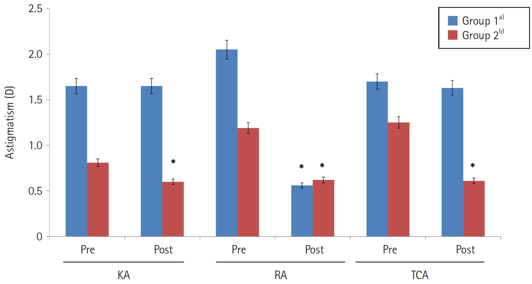
Clinical outcomes were compared between 60 patients who underwent EDOF toric IOL implantation and 60 patients who underwent EDOF IOL implantation combined with LRI. A toric IOL was implanted when corneal astigmatism exceeded 1.0 diopters (D) (group 1), whereas LRI was performed when corneal astigmatism was 1.0 D or less (group 2). Corneal astigmatism, refractive astigmatism, refraction, uncorrected visual acuity, and corrected visual acuity (logMAR) were analyzed in each group before surgery and 3 months postoperatively.
Results
Corneal and total corneal astigmatism did not differ significantly between preoperative and postoperative measurements in group 1 (P>0.05). In group 2, postoperative corneal and total corneal astigmatism (0.60±0.13 and 0.61±0.28 D, respectively) were significantly lower than the corresponding preoperative values (0.81±0.36 and 1.25±0.32 D, respectively; P<0.05). Refractive astigmatism decreased significantly in both group 1 and group 2, from 2.05±0.64 and 1.18±0.41 D preoperatively to 0.56±0.32 and 0.62±0.26 D at 3 months postoperatively, respectively; however, the magnitude of reduction was significantly greater in group 1 than in group 2 (P<0.05). Postoperative refractive astigmatism did not differ significantly between the two groups (P>0.05). In both groups, uncorrected and corrected visual acuity improved significantly after surgery (P<0.05), with no significant between-group difference (P>0.05).
Conclusion
Combining an EDOF IOL with LRI may be effective for mild astigmatism, whereas selecting a toric EDOF IOL may better support visual improvement in patients with moderate to severe astigmatism.
- 226 View
- 2 Download
- Effect of lubricant eye drops administered before visual field testing in patients with glaucoma and dry eye
- Jiyoung Emily Lee, Eun Chul Kim
- Insights Cataract Refract Surg 2026;11(3):91-97. Published online October 29, 2026
- DOI: https://doi.org/10.63375/icrs.26.009
-
Abstract
PDFePub
- Purpose
This study aimed to evaluate the effect of administering lubricant eye drops before visual field testing in patients with glaucoma and dry eye syndrome.
Methods
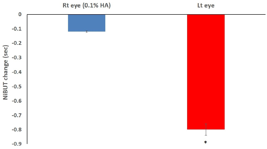
The records of 30 patients (60 eyes) with dry eye syndrome were retrospectively reviewed. Before visual field testing, 0.1% hyaluronic acid eye drops were administered to the right eye, while the left eye received no drops. The efficacy of the lubricant eye drops was assessed by retrospectively comparing tear meniscus height, noninvasive tear film break-up time (NiBUT), and corneal staining scores before and after testing.
Results
Before visual field testing, NiBUT was 5.16±2.19 seconds in the right eye and 5.07±1.98 seconds in the left eye; after testing, it was 5.04±2.36 seconds and 4.27±1.32 seconds, respectively. Tear meniscus height was 0.26±0.11 mm in the right eye and 0.25±0.10 mm in the left eye before testing, decreasing to 0.25±0.11 mm and 0.22±0.10 mm, respectively, afterward. Before testing, corneal staining scores were 1.07±0.74 in the right eye and 1.17±0.86 in the left eye; after testing, they were 1.33±0.76 and 1.90±0.95, respectively. The decreases in NiBUT and tear meniscus height from before to after testing were greater in the left eye than in the right eye, and corneal staining increased significantly (P<0.05). Fixation loss, false-positive, and false-negative rates during visual field testing were all significantly higher in the left eye than in the right eye (P<0.05).
Conclusion
Visual field testing requires patients to keep their eyes open and maintain focus for an extended period, which can exacerbate symptoms in patients with dry eye syndrome and potentially compromise test accuracy. Administering lubricant eye drops before testing, following consultation with an ophthalmologist, is thought to alleviate dry eye symptoms and improve test accuracy.
- 32 View
- 0 Download
Case Reports
- Therapeutic penetrating keratoplasty in rapidly progressive Acanthamoeba keratitis: a case report
- Yunseo Choi, Jaewon Jung, Tae-im Kim
- Insights Cataract Refract Surg 2026;11(3):98-103. Published online July 31, 2026
- DOI: https://doi.org/10.63375/icrs.26.006
-
Abstract
PDFePub
- Purpose
To report the clinical course and postoperative management strategy for emergency therapeutic penetrating keratoplasty (PKP) in a patient with rapidly progressive Acanthamoeba keratitis.
Case
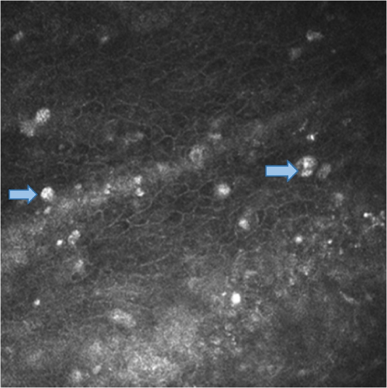
summary: A 23-year-old female patient with a history of long-term orthokeratology lens wear presented with Acanthamoeba keratitis in the right eye. The diagnosis had been established at a referring hospital based on characteristic clinical findings, and topical antiamoebic therapy was initiated, resulting in initial clinical improvement. After polyhexamethylene biguanide was discontinued, an epithelial defect recurred. Amniotic membrane transplantation with adjunctive topical corticosteroids was then performed, after which corneal infiltration and inflammation worsened. At presentation to our clinic, best-corrected visual acuity (BCVA) was light perception, and intraocular pressure was elevated to 34.1 mmHg. Slitlamp biomicroscopic examination revealed extensive corneal infiltration accompanied by hypopyon. Emergency therapeutic PKP was performed the following day because of uncontrolled infection with severe stromal thinning. Postoperatively, intensive antiamoebic therapy was administered, and topical corticosteroids were initially withheld. Systemic immunosuppression was introduced 1 week postoperatively to manage early graft rejection. Management required careful balancing of immunosuppression for graft survival against the risk of recurrent Acanthamoeba infection. Extensive inferior corneal neovascularization was observed and required additional interventions, including subconjunctival bevacizumab injection. At 5 to 6 months postoperatively, the graft remained clear, with no evidence of recurrent infection, and BCVA had improved to 0.7.
Conclusion
Timely emergency therapeutic PKP with careful postoperative management may serve as a vision-preserving strategy in patients with rapidly progressive Acanthamoeba keratitis.
- 748 View
- 4 Download
- Utilization of intraoperative optical coherence tomography during Descemet stripping automated endothelial keratoplasty: a case report
- Ji Hyun Yoon, Ho Sik Hwang
- Insights Cataract Refract Surg 2026;11(3):104-110. Published online October 26, 2026
- DOI: https://doi.org/10.63375/icrs.26.004
-
Abstract
PDFePub
- Purpose
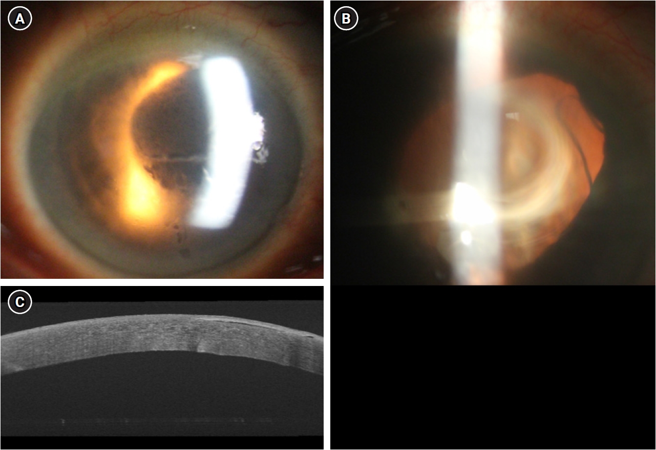
We report a case demonstrating the clinical utility of real-time intraoperative anterior-segment optical coherence tomography (iOCT) for visualizing interface-fluid drainage through venting incisions during Descemet stripping automated endothelial keratoplasty (DSAEK) in a patient with bullous keratopathy.
Case
summary: A 77-year-old male patient with a history of bilateral cataract surgery presented with a several-month history of decreased vision in the right eye. Slit-lamp examination revealed corneal edema with bullae, and central corneal thickness was 700 μm. The endothelial cell count could not be measured by specular microscopy. With a diagnosis of pseudophakic bullous keratopathy, DSAEK was performed. After graft insertion and anterior-chamber air tamponade, iOCT identified residual interface fluid between the host stroma and donor graft. Venting incisions were then created, and the residual fluid was expressed with a sponge. Real-time iOCT confirmed the gradual disappearance of the interface fluid and complete apposition of the graft to the host cornea. After surgery, the graft remained stably attached without rejection or detachment during 3 months of follow- up.
Conclusion
During DSAEK, iOCT allows immediate verification of interface-fluid removal through venting incisions and may help improve graft positioning and early adherence.
- 39 View
- 0 Download
- Recurrent keratoconjunctivitis in X-linked agammaglobulinemia: a microbiome analysis: a case report
- Bo Ree Kim, Kyung Eun Han
- Insights Cataract Refract Surg 2026;11(3):111-116. Published online October 29, 2026
- DOI: https://doi.org/10.63375/icrs.26.007
-
Abstract
PDFePub
- Purpose
To describe recurrent keratoconjunctivitis in a patient with X-linked agammaglobulinemia (XLA) and report the findings of microbiome analysis.
Case
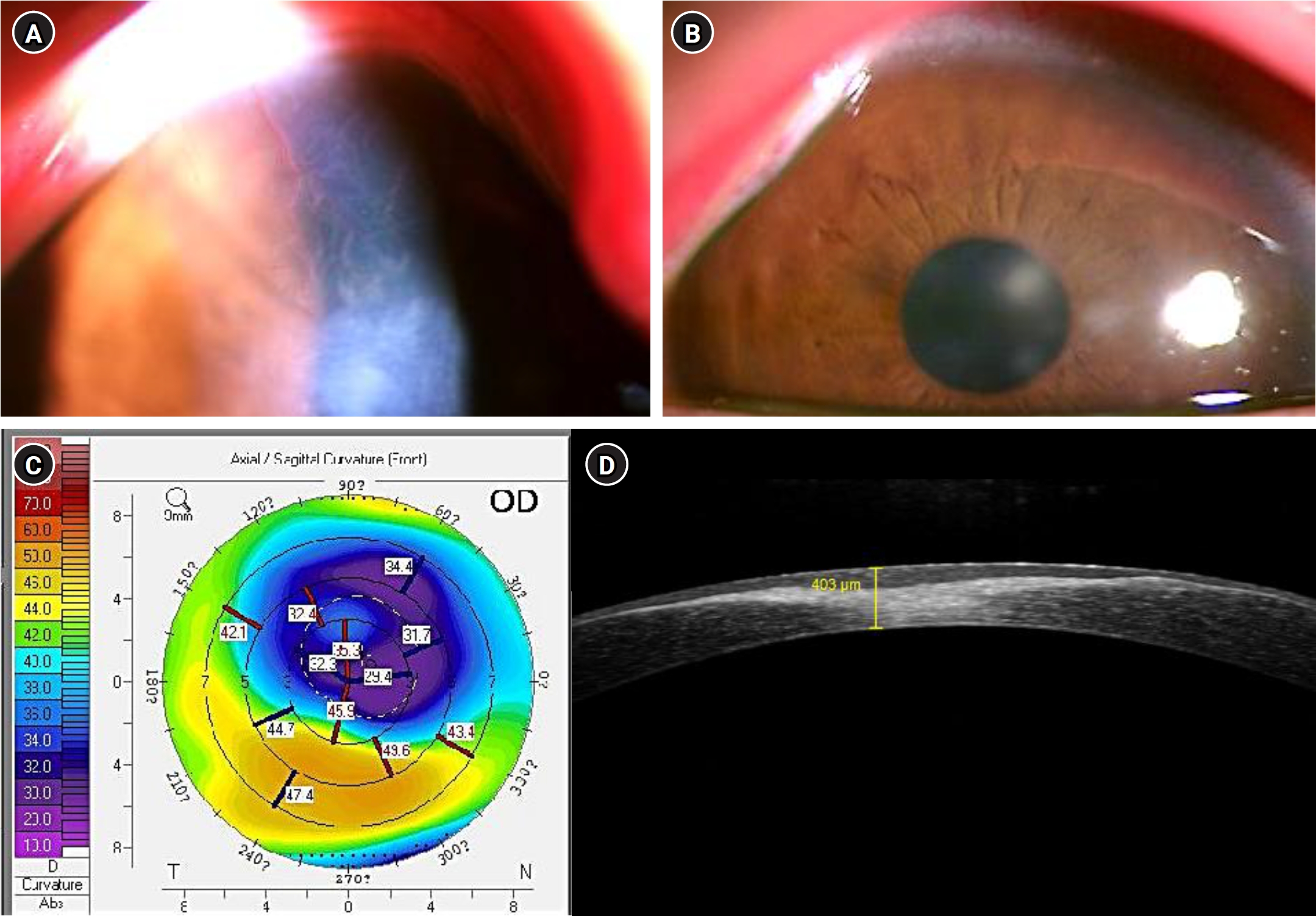
summary: A 15-year-old male with XLA presented with photophobia, epiphora, redness, and reduced vision in the right eye. Examination revealed corneal infiltration, neovascularization (NV), and thinning in the right eye, with multiple subepithelial opacities in the left. The corneal lesions improved transiently with topical corticosteroids and antibiotics but repeatedly worsened approximately 1 week before each scheduled monthly intravenous immunoglobulin (IVIG) infusion. Approximately 23 months later, corneal NV, central opacity, and thinning also developed in the left eye. Cultures of both conjunctivae yielded Haemophilus influenzae; a right eyelid culture also yielded Staphylococcus epidermidis. Conjunctival microbiome analysis confirmed H. influenzae and detected additional organisms—Mycoplasma amphoriforme, Streptococcus pneumoniae, and Staphylococcus aureus.
Conclusion
In this patient with XLA, topical therapy provided only transient improvement in recurrent keratoconjunctivitis. Exacerbations were temporally associated with the presumed trough period before each scheduled IVIG infusion. Conjunctival microbiome analysis detected additional organisms not identified by bacterial culture.
- 33 View
- 1 Download
 First
 Prev



